CROSS REFERENCE TO RELATED APPLICATIONS
-
The present application claims the priority benefit of
US Provisional Patent Application No. 61/246,079, filed September 25, 2009 and US Provsional Patent Application No.
61/306,324, filed February 19, 2010 , each of which is incorporated by reference in its entirety.
FIELD
-
The present invention relates to methods of screening for polypeptide binding agents, e.g. antibodies, that exhibit the ability to kinetically modulate the binding and signaling of biological signaling complexes, e.g., receptor-ligand complexes. The invention also relates to specific polypeptide binding agents characterized by desired kinetic modulating properties.
APPENDIX A
-
This application includes a table, Appendix A, "41726_SecretedProteins.txt", 255 KB in size, created 25 September 2009, submitted with this application.
BACKGROUND
-
Most antibody drugs are conventionally identified by screening for antibodies that bind to either a cell-surface receptor or its cognate ligand, and identifying antibodies that specifically block or stimulate the receptor signaling activity. Many antibody drugs block signaling pathways by binding to either the ligand or receptor, thereby eliminating the ability of the ligand to bind to and activate the receptor. Such blocking antibodies mediate their effect stoichiometrically by preventing the formation of receptor-ligand complex. Conversely, some antibody drugs bind to and activate signaling of a receptor. Such agonist antibodies may mediate their effects by mimicking the natural activity of the ligand and thus do not require the presence of ligand to activate signaling.
SUMMARY
-
The invention provides novel categories of polypeptide binding agents, designated "kinetic modulating drugs" or "kinetic modulators," that have desirable properties for modulating, either positively or negatively, cellular pathway activity involving a target and its signaling partner. The target and/or its signaling partner may be an endogenous or exogenous compound, either proteinaceous or non-proteinaceous in nature, but which optionally may exclude ions and salts. The invention also provides novel methods of identifying such kinetic modulators, based on their effect on binding kinetics between the target and its signaling partner, or based on differential binding of the kinetic modulator for the target (and/or its signaling partner) in complexed form versus uncomplexed form. The polypeptide binding agent may bind the target, its signaling partner and/or a complex comprising the target and its signaling partner. This discovery allows biophysical screening assays to be designed which can identify modulators of cellular pathway activity suitable for therapeutic use.
-
In some aspects, assays are provided to identify polypeptide binding agents which modulate the binding kinetics between a target and its signaling partner. Nonlimiting examples of targets include, e.g. a secreted protein of any of the Accession nos. as set forth in Appendix A (or SEQ ID NOS: 1-88). These secreted proteins include a number of secreted membrane-bound receptors. Appendix A herewith lists human secreted proteins as compiled by the Swissprot/EMBL database (see e.g.,
Boeckmann et al. "The SWISS-PROT protein knowledgebase and its supplement TrEMBL in 2003", Nucleic Acids Res. 31:365-370(2003). Appendix A sets out the Swissprot accession number for the amino acid sequence of the secreted protein, the name of the protein (and all acronyms or related names) and the length of the amino acid sequence in the database. As used herein, a "signaling partner" is a binding partner of a target that, when bound to the target, forms a signaling complex or is part of a signaling complex that activates or inhibits a cellular pathway. The presence of such kinetic modulator polypeptide binding agents alters (strengthens or weakens) the apparent binding affinity of the target for its signaling partner, thus altering the dose-response of the target for activating the cellular pathway. Alternatively, a kinetic modulator polypeptide binding agent that alters (increases or decreases) the on-rate or alters (decreases or increases) the off-rate of the target for its signaling partner can also change (increase or decrease) the residency time of the target complexed with the signaling partner, change the rate of receptor internalization and/or change the degree of phosphorylation of signaling proteins that are activated or deactivated by the signaling partner complex. Such changes could significantly alter the relative activation of different signaling pathways by the complexation of target and signaling partner and thus alter the dose-response of the target for activating the cellular pathway. Such kinetic modulators are expected to have advantages over conventional therapeutic drugs, including improved safety profiles, altered clearance rates, broader therapeutic windows and less frequent dosing. Where the target is an exogenous compound that is being administered to the patient, administration of the kinetic modulator as an adjunct therapy with the target can alter (e.g., decrease) the total amount (daily, weekly or monthly) and/or the frequency of dosing of the target.
-
The invention provides methods of identifying candidate kinetic modulating drugs that are polypeptide binding agents, excluding traditional small molecule drugs such as non-polymeric organic chemical compounds having a molecular weight of about 1000 daltons or less. Examples of specifically contemplated polypeptide binding agents include antibodies, including antigen-binding fragments thereof, peptibodies, polypeptides and peptides, optionally conjugated to other peptide moieties or non-peptidic moieties. Examples of antibodies include monoclonal antibodies, tetrameric immunoglobulins comprising two heavy chains and two light chains, single chain antibodies, single domain antibodies, antibody fragments, scFv, Fab, CDRs, rodent antibodies, mammalian antibodies, human antibodies, chimeric antibodies and humanized antibodies.
-
The invention provides methods of identifying a candidate polypeptide binding agent, e.g. an antibody, that modulates binding between first and second components of a signaling complex (the target and signaling partner, or vice versa). Examples of such first and/or second components include any of the secreted proteins of Appendix A (or SEQ ID NOS: 1-88) and endogenous or exogenous signaling partners of such secreted proteins, or any of the ligands or receptors or transmembrane proteins described herein. In some embodiments, the first and second components are polypeptides. In exemplary specific embodiments, the first and second components are endogenous.
-
In one aspect, the methods of identifying a candidate kinetic modulating drug include (a) measuring a binding affinity or binding rate parameter of said first component for said second component, in the presence of a test polypeptide binding agent, e.g. antibody, (b) measuring a binding affinity or binding rate parameter of said first component for said second component in the absence of said test polypeptide binding agent; and (c) identifying said test polypeptide binding agent as a candidate kinetic modulating drug when said test polypeptide binding agent exhibits at least a 1.5-fold difference in a binding affinity or binding rate parameter measured in steps (a) and (b). Figure 1 shows a schematic diagram illustrating some exemplary embodiments. In some embodiments, the difference in binding affinity or binding rate parameter ranges from about 1.5-fold (i.e., 50%) to, optionally, about 1000-fold, or about 1.5-fold to about 100-fold, or about 2-fold to 25-fold, or about 2-fold to 50-fold, or about 1.5-fold to about 25-fold, or about 1.5-fold to about 50-fold, e.g. at least 1.5-fold, 2-fold, 3-fold, 4-fold, 5-fold, 6-fold, 7-fold, 8-fold, 9-fold, 10-fold, 11-fold, 12-fold, 13-fold, 14-fold, 15-fold, 16-fold, 17-fold, 18-fold, 19-fold or 20-fold, or up to 500-fold, or up to 200-fold, or up to 150-fold, or up to 100-fold, or up to 90-fold, or up to 80-fold, or up to 70-fold, or up to 60-fold, or up to 50-fold, or up to 40-fold, or up to 30-fold. In some embodiments, the test polypeptide binding agent is identified as a candidate positive modulator if the test polypeptide agent strengthens a binding affinity or binding rate parameter between said first component and said second component (e.g., reduced KD, or increased KA, or reduced ratio of off-rate/on-rate, or increased ratio of on-rate/off-rate, or increased on-rate, or decreased off-rate). In other embodiments, the test polypeptide agent is identified as a candidate negative modulator if the test polypeptide agent weakens a binding affinity or binding rate parameter between said first component and said second component (e.g., increased KD, or decreased KA, or increased ratio of off-rate/on-rate, or decreased ratio of on-rate/off-rate, or decreased on-rate, or increased off-rate).
-
In some alternative embodiments, where a stronger binding rate parameter (e.g., increased association or residency time, via increased on-rate or decreased off-rate) results in increased relative activation of the desired signaling pathway, even when binding affinity is not detectably changed, the test polypeptide binding agent is identified as a candidate positive modulator by identifying the desired-fold strengthening in binding rate parameter. Where a weaker binding rate parameter (e.g., decreased association or residency time, via decreased on-rate or increased off-rate) results in increased relative activation of the desired signaling pathway, even when binding affinity is not detectably changed, the test polypeptide binding agent is identified as a candidate positive modulator by identifying the desired-fold weakening in binding rate parameter. Similarly, where a stronger binding rate parameter (e.g., increased association or residency time, via increased on-rate or decreased off-rate) results in decreased relative activation of the desired signaling pathway, even when binding affinity is not detectably changed, the test polypeptide binding agent is identified as a candidate negative modulator by identifying the desired-fold strengthening in binding rate parameter. Where a weaker binding rate parameter (e.g., decreased association or residency time, via decreased on-rate or increased off-rate) results in decreased relative activation of the desired signaling pathway, even when binding affinity is not detectably changed, the test polypeptide binding agent is identified as a candidate negative modulator by identifying the desired-fold weakening in binding rate parameter.
-
In another aspect, the methods of identifying a candidate kinetic modulating drug include (a) (i) measuring a binding affinity or binding rate parameter of a test polypeptide binding agent, e.g. antibody, for said first component in the presence of said second component, or (ii) measuring a binding affinity or binding rate parameter of a test polypeptide binding agent for said second component in the presence of said first component; and (b) (i) measuring a binding affinity or binding rate parameter of said test polypeptide binding agent for said first component in the absence of said second component, or (ii) measuring a binding affinity or binding rate parameter of said test polypeptide binding agent for said second component in the absence of said first component; and (c) identifying said test polypeptide binding agent as a candidate kinetic modulating drug when said test polypeptide binding agent exhibits a 1.5-fold to 100-fold difference in the binding affinity or binding rate parameter measured in steps (a) and (b). Figure 2 shows a schematic diagram illustrating some exemplary embodiments, in which interaction is measured in the presence and absence of the second component.
-
In some embodiments, the difference in binding affinity or binding rate parameter measured in steps (a) and (b) ranges from about 1.5-fold (i.e., 50%) to, optionally, about 1000-fold, or about 1.5-fold to about 100-fold, or about 2-fold to 25-fold, or about 2-fold to about 50-fold, or about 1.5-fold to about 25-fold, or about 1.5-fold to about 50-fold, e.g. at least 1.5-fold, 2-fold, 3-fold, 4-fold, 5-fold, 6-fold, 7-fold, 8-fold, 9-fold, 10-fold, 11-fold, 12-fold, 13-fold, 14-fold, 15-fold, 16-fold, 17-fold, 18-fold, 19-fold or 20-fold, or up to 500-fold, or up to 200-fold, or up to 150-fold, or up to 100-fold, or up to 90-fold, or up to 80-fold, or up to 70-fold, or up to 60-fold, or up to 50-fold, or up to 40-fold, or up to 30-fold. In some embodiments, the test polypeptide binding agent is identified as a candidate positive modulator if the binding affinity or binding rate parameter measured in step (a) is stronger than the binding affinity or binding rate parameter measured in step (b). In other embodiments, the test polypeptide binding agent is identified as a candidate negative modulator if the binding affinity or binding rate parameter measured in step (b) is stronger than the binding affinity or binding rate parameter measured in step (a).
-
Any of the foregoing methods can be carried out as high throughput assays, in which multiple polypeptide binding agents (e.g., at least 5, 10, 15, 20, 25, 30, 35, 40, 50, 100, 150, 200, 500, 1,000, 10,000, or 25,000) are screened simultaneously or sequentially. In some embodiments, the methods further involve assaying a plurality of test polypeptide binding agents, e.g. antibodies, for binding affinity to any one of (a) the first component, (b) the second component, or (c) a complex comprising the first component and second component, optionally prior to measuring differences in binding affinity or binding rate parameter. Such prescreening of libraries can also be carried out as high throughput assays, in which multiple polypeptide binding agents (e.g., at least 5, 10, 15, 20, 25, 30, 35, 40, 50, 100, 150, 200, 500 or 1000) are screened simultaneously or sequentially. In some embodiments, the plurality of test polypeptide binding agents screened are variants of a parent polypeptide binding agent made by introducing one or more different mutations into a parent polypeptide binding agent.
-
In further embodiments, the polypeptide binding agents may be screened for selectivity of effect for the first or second component, compared to a different binding partner such as a decoy receptor, clearance receptor, or alternate signal pathway component. Such methods may involve identifying a polypeptide binding agent that does not significantly change the binding affinity or binding rate parameter of the first or second component for a different binding partner, such binding partner being neither the first nor second component.
-
Any of the preceding measurements of binding affinity or binding rate parameter may be carried out in assays where one or more of the first component, second component and polypeptide binding agent are in solution, or in assays where one or more of the first component, second component and polypeptide binding agent are linked to a solid phase (covalently or noncovalently), or in assays where one or more of the first component, second component and polypeptide binding agent are expressed on a cell surface. The first and/or second components may each themselves be complexes of multiple compounds. The first and/or second components (e.g., target or signaling partner or vice versa) may be soluble or membrane-bound ligands or receptors, including but not limited to 7-transmembrane receptors, G-protein coupled receptors (GPCRs), adrenergic receptors, neurotransmitter receptors, olfactory receptors, opioid receptors, chemokine receptors, rhodopsin, receptor tyrosine kinases, growth factor receptors, integrins, and toll-like receptors, enzymes, or substrates.
-
Any of the preceding methods may further include recloning and expressing, or synthesizing and expressing, or synthesizing, the candidate kinetic modulating polypeptide binding agent; purifying and/or sequencing the kinetic modulator; adding or replacing an Fc region or fragment thereof; formulating the kinetic modulator or a variant, e.g. an antibody comprising at least three or six of the same CDRs of the parent antibody, in a sterile composition with a sterile pharmaceutically acceptable diluent; and/or administering the kinetic modulator or a variant to an animal.
-
Any of the preceding methods may further include measuring the level of signaling mediated by the signaling complex in the presence and absence of the test polypeptide binding agent, and determining whether the test polypeptide binding agent is additionally an agonist, partial agonist, antagonist or partial antagonist. In certain embodiments, the agonist or partial agonist is an allosteric agonist.
-
In related aspects, the invention provides a polypeptide binding agent, e.g. an antibody, identified by any of the preceding methods or any of the methods described elsewhere herein.
-
In a separate aspect, the invention also provides polypeptide binding agents with desired characteristics. In some embodiments, the invention provides a positive modulator that (a) binds to the target, e.g., a secreted protein of any of Appendix A (or SEQ ID NOS: 1-88) with an equilibrium dissociation constant KD of about 10-5M or less, e.g., 10-6M or less, or 10-7M or less, or 10-8M or less (wherein a lower number indicates higher binding affinity), and (b) is capable of improving the binding affinity KD between said target and its signaling partner by about 1.5-fold (i.e., 50%) to, optionally, about 1000-fold, or about 1.5-fold to about 100-fold, or about 2-fold to 25-fold, or about 2-fold to about 50-fold, or about 1.5-fold to about 25-fold, or about 1.5-fold to about 50-fold, e.g. at least 1.5-fold, 2-fold, 3-fold, 4-fold, 5-fold, 6-fold, 7-fold, 8-fold, 9-fold, 10-fold, 11-fold, 12-fold, 13-fold, 14-fold, 15-fold, 16-fold, 17-fold, 18-fold, 19-fold or 20-fold, or up to 500-fold, or up to 200-fold, or up to 150-fold, or up to 100-fold, or up to 90-fold, or up to 80-fold, or up to 70-fold, or up to 60-fold, or up to 50-fold, or up to 40-fold, or up to 30-fold. In other embodiments, the invention provides a negative modulator that (a) binds to the target, e.g., secreted protein of any of Appendix A (or SEQ ID NOS: 1-88) with an equilibrium dissociation constant KD of about 10-5M or less, e.g., 10-6M or less, or 10-7M or less, or 10-8M or less, and (b) is capable of reducing the binding affinity KD between said target and its signaling partner by about 1.5-fold (i.e., 50%) to, optionally, about 1000-fold, or about 1.5-fold to about 100-fold, or about 2-fold to 25-fold, or about 2-fold to about 50-fold, or about 1.5-fold to about 25-fold, or about 1.5-fold to about 50-fold, e.g. at least 1.5-fold, 2-fold, 3-fold, 4-fold, 5-fold, 6-fold, 7-fold, 8-fold, 9-fold, 10-fold, 11-fold, 12-fold, 13-fold, 14-fold, 15-fold, 16-fold, 17-fold, 18-fold, 19-fold or 20-fold, or up to 500-fold, or up to 200-fold, or up to 150-fold, or up to 100-fold, or up to 90-fold, or up to 80-fold, or up to 70-fold, or up to 60-fold, or up to 50-fold, or up to 40-fold, or up to 30-fold.
-
Any of such polypeptide binding agents may be further subject to purification, to obtain a substantially homogeneous composition, e.g. at least about 90%, 95%, 97%, 98%, 99% or 99.5% pure.
-
The invention further provides methods of preparing a sterile pharmaceutical composition comprising adding a sterile pharmaceutically acceptable diluent to such polypeptide binding agents, sterile compositions of such polypeptide binding agents, e.g., in a therapeutically effective amount, and methods of administering such sterile compositions, e.g. to modulate (increase or decrease) signaling of a complex comprising the secreted protein.
-
It is understood that each feature or embodiment, or combination, described herein is a non-limiting, illustrative example of any of the aspects of the invention and, as such, is meant to be combinable with any other feature or embodiment, or combination, described herein. For example, where features are described with language such as "one embodiment", "some embodiments", "further embodiment", "specific exemplary embodiments", and/or "another embodiment", each of these types of embodiments is a non-limiting example of a feature that is intended to be combined with any other feature, or combination of features, described herein without having to list every possible combination. Such features or combinations of features apply to any of the aspects of the invention. Similarly, where a method describes identifying polypeptide binding agents, such as antibodies, characterized by certain features, polypeptide binding agents characterized by those features are also contemplated by the invention. Where examples of values falling within ranges are disclosed, any of these examples are contemplated as possible endpoints of a range, any and all numeric values between such endpoints are contemplated, and any and all combinations of upper and lower endpoints are envisioned.
-
Numerous additional aspects and advantages of the invention will become apparent to those skilled in the art upon consideration of the following detailed description of the invention which describes presently preferred embodiments thereof. All U.S. patents, U.S. patent application publications, U.S. patent applications, foreign patents, foreign patent applications, and non-patent publications referred to in this application, are incorporated herein by reference, in their entireties.
BRIEF DESCRIPTION OF THE FIGURES
-
- Figure 1 is a schematic diagram to illustrate measurement of binding performed in the presence or absence of test polypeptide binding agent.
- Figure 2 is a schematic diagram to illustrate measurement of binding performed in the presence or absence of a second complex component.
- Figure 3 shows the predicted effects of kinetic modulating antibodies on signaling activity at (A) varying ligand concentrations; (B) varying modulator concentrations (non-agonist antibody); and (C) varying modulator concentrations (agonist antibody).
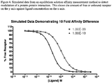
- Figure 4 shows simulated data from an equilibrium solution affinity measurement method to detect modulation of a protein-protein interaction.
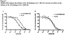
- Figure 5 shows the effects of XOMA 052 on the affinity of IL-1β binding to IL-1 sRI (A), and to IL-1 sRII (B).
- Figure 6 shows (A) neutralization of IL-1β activity by XOMA 052 at the EC50 of IL-1β for the cell assay, and (B, C) that negative affinity modulation of the IL-1β to IL-1 sRI interaction results in an altered cellular dose-response to IL-1β resulting in an increase in the IC50.
- Figure 7 shows the amount of total IL-1β remaining in circulation 48 hours following injection of antibody/ IL-1β complexes.
- Figure 8 is an illustration of the regulation of IL-1β activity by different drug types in T2D.
- Figure 9 shows results from solid phase affinity measurement assays to identify antibodies which modulate the GCSF-GCSFR binding interaction as described in Example 2.
- Figure 10 shows GCSF-dependent binding of A10(B6) antibody to GCSFR-transfected BAF3 cells.
- Figure 11 shows sample results from a cell-based affinity measurement assay to identify antibodies which modulate the hINS-INSR binding interaction.
- Figure 12 shows sample results from a cell-based affinity measurement assay to measuremodulation of the hINS-INSR binding interaction by test antibodies.
- Figure 13 shows example results from an assay measuring the ability of test anti-INSR antibodies to stimulate pIRS-1 phosphorylation.
- Figure 14 is a table showing insulin EC50 values from the pIRS-1 assay in the presence or absence of fixed concentrations of various test antibodies.
- Figure 15 shows blood glucose levels in 20 week old DIO mice fed a high fat diet and treated with partial agonist anti-INSR antibodies: A. Line graph of glucose levels. B. Bar chart of glucose levels showing statistically significant reduction in blood glucose after injection of partial agonist anti-INSR antibody.
- Figure 16 illustrates that administration of a partial agonist anti-INSR antibody improves glycemic control in DIO mice: A. Glucose tolerance test timecourse; B. Fasting blood glucose levels; C. Glucose tolerance test; area under curve (AUC).
- Figure 17 shows that a positive modulator anti-INSR antibody improves insulin sensitivity in DIO mice: A. Insulin tolerance test timecourse; B. Fasting blood glucose levels; C. Insulin tolerance test; area under curve (AUC).
- Figure 18 shows that a positive modulator anti-INSR antibody improves glycemic control in DIO mice: A. Glucose tolerance test timecourse; B. Fasting blood glucose levels; C. Glucose tolerance test; area under curve (AUC).
- Figure 19 illustrates the dose response from a partial allosteric agonist in comparison to the dose response to the endogenous ligand (A) or activation by ligand in the presence or absence of the allosteric agonist (B).
- Figure 20 shows the dose response from a positve allosteric modulator antibody in comparison to the dose response to the endogenous ligand (A) or the dose response of an endogenous ligand in the presence and absence of a positive allosteric modulator antibody (B).
- Figure 21 illustrates the activation parameters for a set of partial allosteric agonists alone relative to the endogenous ligand insulin. Data obtained from measurements of percent Akt phosphorylation at Ser473.
- Figure 22 illustrates the activation properties of insulin in the presence of 10 ug/ml partial allosteric agonist antibodies relative to the maximal response to the endogenous ligand in the presence of a negative control antibody. Data obtained from measurements of percent Akt phosphorylation at Ser473.
DETAILED DESCRIPTION
-
The invention provides kinetic modulating drugs that are polypeptide binding agents, uses thereof, and various methods of identifying kinetic modulating drugs. These kinetic modulators can induce either a positive or negative effect on the cellular response by altering the kinetic rate constants for assembly and dissociation of signaling complex components or by other mechanisms including altering the structural state of the signaling complex, e.g., by binding to a transition state and accelerating the activation of signaling.
-
Modulation of a signaling complex can result in an increase or decrease in sensitivity to signal input and concomitant increases or decreases in signal transduction. Administration of these kinetic modulators increases or decreases the sensitivity of the cellular pathway and/or absolute levels of the cellular response. The kinetic modulators of the invention, depending on their properties, can function as a modulator, potentiator, regulator, effector or sensitizer.
-
Many antibody drugs act to block signaling pathways by binding to either a cell-surface receptor or its cognate ligand and eliminating the ability of the ligand to bind to and activate the receptor. Such blocking drugs mediate their effect stoichiometrically by preventing the formation of receptor-ligand complex. However, most pathways that have been linked to disease when abnormally activated also have normal developmental or homeostatic roles in normal biology. This observation is particularly true for the immune system, where highly potent cytokines such as TNF-α and IL-6 drive inflammation in pathological contexts but also have important beneficial roles in the control of infections. Successful treatment of some diseases may therefore require attenuation rather than complete inhibition of signaling pathways to restore a normal physiological state with acceptable side-effect profiles. The kinetic modulators provided by the invention are expected to provide such advantages.
-
Other therapeutic drugs affect cellular signaling pathways by binding to a cell-surface receptor and altering the activity of the receptor. Such direct agonist drugs may mediate their effects by mimicking the natural activity of the ligand and thus have inherent activity i.e. they do not require the presence of ligand to mediate their effects. Further therapeutic drugs affect cellular signaling pathways by binding to a ligand. Such indirect agonist drugs may mediate their effects by altering ligand stability or valency.
-
Biological processes are generally regulated in a continuous rather than binary manner, and thus in many cases modulation of pathway activity may be a more appropriate therapeutic strategy than complete pathway blockade or stimulation. Performing functional, cell-based screens for modulation of pathway activity, rather than for complete pathway blockade or stimulation,is laborious and may not readily be readily performed in a high throughput manner, since such screens generally require a known concentration of test compound and may be sensitive to any impurities in the test compound preparation. In particular, the ability to perform high throughput functional, cell-based screens for modulation of pathway activity is restricted for cell-impermeable molecules which are unable to enter the intracellular environment, and especially for recombinant biological molecules which may have different expression levels, degrees of purity and stability in the production system used. In addition, some binding interactions may have no signaling output to measure in a functional screen (e.g. in the case of decoy receptors, decoy substrates, or inactive forms of a target) making it difficult to identify agents that perturb these interactions.
-
The present invention overcomes these disadvantages and provides a means for identifying positive and negative kinetic modulators of the desired activity and desired potency in a high throughput manner.
Definitions
-
The term "compound" refers to any chemical compound, organic or inorganic, endogenous or exogenous, including, without limitation, polypeptides, proteins, peptides, small molecules, nucleic acids (e.g. DNA and RNA), carbohydrates, lipids, fatty acids, steroids, purines, pyrimidines, peptidomimetics, polyketides and derivatives, structural analogs or combinations thereof. "Endogenous" means naturally occurring in a mammal, while "exogenous" means not naturally occurring in the mammal, e.g. an administered foreign compound.
-
The term "polypeptide binding agent" refers to a polypeptide that is capable of specifically binding an antigen, e.g. a target or its signaling partner, or that is capable of binding an antigen with a measurable binding affinity. Examples of polypeptide binding agents include antibodies, peptibodies, polypeptides and peptides, optionally conjugated to other peptide moieties or non-peptidic moieties. Antigens to which a polypeptide binding agent may bind include any proteinaceous or non-proteinaceous molecule that is capable of eliciting an antibody response, or that is capable of binding to a polypeptide binding agent with detectable binding affinity greater than non-specific binding. The antigen to which a kinetic modulating polypeptide binding agent binds may include a target, a signaling partner of a target, and/or a complex comprising the target and its signaling partner.
-
The term "antibody" is used in the broadest sense and includes fully assembled antibodies, tetrameric antibodies, monoclonal antibodies, polyclonal antibodies, multispecific antibodies (e.g., bispecific antibodies), antibody fragments that can bind an antigen (e.g., Fab', F'(ab)2, Fv, single chain antibodies, diabodies), and recombinant peptides comprising the forgoing as long as they exhibit the desired biological activity. An "immunoglobulin" or "tetrameric antibody" is a tetrameric glycoprotein that consists of two heavy chains and two light chains, each comprising a variable region and a constant region. Antigen-binding portions may be produced by recombinant DNA techniques or by enzymatic or chemical cleavage of intact antibodies. Antibody fragments or antigen-binding portions include, inter alia, Fab, Fab', F(ab')2, Fv, domain antibody (dAb), complementarity determining region (CDR) fragments, single-chain antibodies (scFv), single chain antibody fragments, chimeric antibodies, diabodies, triabodies, tetrabodies, minibody, linear antibody; chelating recombinant antibody, a tribody or bibody, an intrabody, a nanobody, a small modular immunopharmaceutical (SMIP), a antigen-binding-domain immunoglobulin fusion protein,a camelized antibody, a VHH containing antibody, or a variant or a derivative thereof, and polypeptides that contain at least a portion of an immunoglobulin that is sufficient to confer specific antigen binding to the polypeptide, such as one, two, three, four, five, or six CDR sequences, as long as the antibody retains the desired biological activity.
-
"Monoclonal antibody" refers to an antibody obtained from a population of substantially homogeneous antibodies, i.e., the individual antibodies comprising the population are identical except for possible naturally occurring mutations that may be present in minor amounts.
-
"Antibody variant" as used herein refers to an antibody polypeptide sequence that contains at least one amino acid substitution, deletion, or insertion in the variable region of the natural antibody variable region domains. Variants may be substantially homologous or substantially identical to the unmodified antibody.
-
A "chimeric antibody," as used herein, refers to an antibody containing sequence derived from two different antibodies (see, e.g.,
U.S. Patent No. 4,816,567 ) which typically originate from different species. Most typically, chimeric antibodies comprise human and rodent antibody fragments, generally human constant and mouse variable regions.
-
A "neutralizing antibody" is an antibody molecule which is able to eliminate or significantly reduce a biological function of an antigen to which it binds. Accordingly, a "neutralizing" antibody is capable of eliminating or significantly reducing a biological function, such as enzyme activity, ligand binding, or intracellular signaling.
-
An "isolated" antibody is one that has been identified and separated and recovered from a component of its natural environment. Contaminant components of its natural environment are materials that would interfere with diagnostic or therapeutic uses for the antibody, and may include enzymes, hormones, and other proteinaceous or non-proteinaceous solutes. In preferred embodiments, the antibody will be purified (1) to greater than 95% by weight of antibody as determined by the Lowry method, and most preferably more than 99% by weight, (2) to a degree sufficient to obtain at least 15 residues of N-terminal or internal amino acid sequence by use of a spinning cup sequenator, or (3) to homogeneity by SDS-PAGE under reducing or nonreducing conditions using Coomassie blue or, preferably, silver stain. Isolated antibody includes the antibody in situ within recombinant cells since at least one component of the antibody's natural environment will not be present. Ordinarily, however, isolated antibody will be prepared by at least one purification step.
-
As used herein, an antibody that "specifically binds" is "antigen specific", is "specific for" antigen or is "immunoreactive" with an antigen refers to an antibody or polypeptide binding agent of the invention that binds an antigen with greater affinity than other antigens of similar sequence. In one aspect, the polypeptide binding agents of the invention, or fragments, variants, or derivatives thereof, will bind with a greater affinity to human antigen as compared to its binding affinity to similar antigens of other, i.e., non-human, species, but polypeptide binding agents that recognize and bind orthologs of the target are within the scope of the invention.
-
For example, a polypeptide binding agent that is an antibody or fragment thereof "specific for" its cognate antigen indicates that the variable regions of the antibodies recognize and bind the desired antigen with a detectable preference (e.g., where the desired antigen is a polypeptide, the variable regions of the antibodies are able to distinguish the antigen polypeptide from other known polypeptides of the same family, by virtue of measurable differences in binding affinity, despite the possible existence of localized sequence identity, homology, or similarity between family members). It will be understood that specific antibodies may also interact with other proteins (for example, S. aureus protein A or other antibodies in ELISA techniques) through interactions with sequences outside the variable region of the antibodies, and in particular, in the constant region of the molecule. Screening assays to determine binding specificity of a polypeptide binding agent, e.g. antibody, for use in the methods of the invention are well known and routinely practiced in the art. For a comprehensive discussion of such assays, see Harlow et al. (Eds), Antibodies A Laboratory Manual; Cold Spring Harbor Laboratory; Cold Spring Harbor, NY (1988), . Antibodies for use in the invention can be produced using any method known in the art.
-
The term "epitope" refers to that portion of any molecule capable of being recognized by and bound by a selective binding agent at one or more of the antigen binding regions. Epitopes usually consist of chemically active surface groupings of molecules, such as, amino acids or carbohydrate side chains, and have specific three-dimensional structural characteristics as well as specific charge characteristics. Epitopes as used herein may be contiguous or non-contiguous.
-
The term "derivative" when used in connection with polypeptide binding agents and polypeptides of the invention refers to polypeptides chemically modified by such techniques as ubiquitination, conjugation to therapeutic or diagnostic agents, labeling (e.g., with radionuclides or various enzymes), covalent polymer attachment such as pegylation (derivatization with polyethylene glycol) and insertion or substitution by chemical synthesis of amino acids such as ornithine, which do not normally occur in human proteins. Derivatives retain the binding properties of underivatized molecules of the invention.
-
"Detectable moiety" or a "label" refers to a composition detectable by spectroscopic, photochemical, biochemical, immunochemical, or chemical means. For example, useful labels include 32P, 35S, fluorescent dyes, electron-dense reagents, enzymes (e.g., as commonly used in an ELISA), biotin-streptavadin, dioxigenin, haptens and proteins for which antisera or monoclonal antibodies are available, or nucleic acid molecules with a sequence complementary to another labeled nucleic acid molecule. The detectable moiety often generates a measurable signal, such as a radioactive, chromogenic, or fluorescent signal, that can be used to quantitate the amount of bound detectable moiety in a sample.
-
"Peptides" or "oligopeptides" are short amino acid sequences, typically between 3 and 100 amino acid residues in length and encompass naturally occurring amino acid residues and non-naturally occurring analogs of residues which may be used singly or in combination with naturally occurring amino acid residues in order to give the peptide a particular conformational specificity or a particular biological activity, such as resistance to proteolysis. Peptides include repeats of peptide sequences and may include 2, 3, 4, 5, 6, 7, 8, 9, 10 or more copies of an amino acid sequence arranged head-to-tail or head-to-head. Peptides may be conjugated to non-peptidic moieties, e.g. [expand]. Peptides include dimers, trimers or higher order multimers, e.g. formed through conjugation to other polymeric or non-polymeric moieties, such as PEG.
-
"Polypeptides" are longer amino acid sequences, typically 100 or more amino acid residues in length, and encompass naturally occurring amino acid residues and non-naturally occurring analogs of residues which may be used singly or in combination with naturally occurring amino acid residues in order to give the polypeptide a particular conformational specificity or a particular biological activity, such as resistance to proteolysis.
-
As used herein, a "peptibody" is a fusion polypeptide comprising one or more peptides fused to all or a portion of an immunoglobulin (Ig) constant region. See, e.g.,
U.S. Pat. No. 6,660,843 . The peptide may be any naturally occurring or recombinantly prepared or chemically synthesized peptide that binds to the antigen. The peptide may be repeated and may include 2, 3, 4, 5, 6, 7, 8, 9, 10 or more copies of an amino acid sequence arranged head-to-tail or head-to-head. The portion of the Ig constant region may include at least one constant region domain (e.g., CH1, CH2, CH3, and/or CH4), multiple domains (e.g., CH2 with CH3), multiple copies of domains (e.g., CH2-CH2), any fragment of a constant domain that retains the desired activity, e.g. the salvage receptor epitope responsible for the prolonged half-life of immunoglobulins in circulation, or any combinations thereof.
-
A "small" molecule or "small" organic molecule is defined herein as a non-polymeric organic chemical compound having a molecular weight of about 1000 Daltons or less.
-
As used herein, a "signaling complex" is an assembly of proteins and/or endogenous or exogenous compounds that mediate the transduction of a cellular signal. Examples of a signaling complex include, but are not limited to, a ligand bound to a membrane bound receptor, an enzyme bound to a substrate or any cellular molecules that associate to propagate biochemical reactions that are involved in a signal cascade. Signaling complexes can also include coreceptors, cofactors, scaffold proteins, allosteric modulators and numerous other types of proteins and molecules that are involved in cellular signal transduction. Signaling complexes can be formed transiently or can be long lived. The molecular constituents or components of a signaling complex can vary over time and can be dependent on activation state of each component and the cellular environment. Signaling complexes can undergo chemical modification and regulation that can induce a spectrum of effects on the complex including subtle changes in transduction activity, complete inactivation and constitutive activation or both positive and negative modulation. A component of a signaling complex may be a protein, e.g. a secreted protein of any of Appendix A (or SEQ ID NOS: 1-88), that can exist in association with other proteins and/or compounds in a complex ("complexed") or separately therefrom ("uncomplexed").
-
The term "therapeutically effective amount" is used herein to indicate the amount of kinetic modulator composition of the invention that is effective to ameliorate or lessen symptoms or signs of disease associated with abnormal (e.g. abnormally high or abnormally low) signaling of the signaling complex.
-
As used herein "binding" is the physical association between two or more distinct molecular entities that results from a specific network of non-covalent interactions consisting of one or more of the weak forces including hydrogen bonds, Van der Waals, ion-dipole and hydrophobic interactions and the strong force ionic bonds. The level or degree of binding may be measured in terms of affinity. Affinity, or "binding affinity", is a measure of the strength of the binding interaction between two or more distinct molecular entities that can be defined by equilibrium binding constants or kinetic binding rate parameters. Examples of suitable constants or parameters and their measurement units are well known in the art and include but are not limited to equilibrium association constant (KA), e.g. about 105M-1 or higher, about 106M-1 or higher, about 107M-1 or higher, about 108M-1 or higher, about 109M-1 or higher, about 1010M-1 or higher, about 1011M-1 or higher or about 1012M-1 or higher; equilibrium dissociation constant (KD), e.g., about 10-5M or less, or about 10-6M or less, or about 10-7M or less, or about 10-8M or less, or about 10-9M or less, or about 10-10M or less, or about 10-11M or less, or about 10-12M or less; on-rate (e.g., sec-1, mol-1) and off-rate (e.g., sec-1)). In the case of KA, higher values mean "stronger" or "strengthened" binding affinity while in the case of KD, lower values mean "stronger" or "strengthened" binding affinity. As used herein, a "strengthened" binding rate parameter means increased residency time, faster association or slower dissociation. As used herein, a "weakened" binding rate parameter means decreased residency time, slower association or faster dissociation. In the case of on-rate, higher values mean faster or more frequent association and thus generally result in strengthened binding affinity. In the case of off-rate, lower values generally mean slower dissociation and thus generally result in stronger binding affinity. However, it is the ratio of the on-rate and off-rate that indicates binding affinity, as explained in further detail later.
-
Affinity between two compounds, e.g. between an antibody and an antigen, or between first and second components of a signaling complex, may be measured directly or indirectly. Indirect measurement of affinity may be performed using surrogate properties that are indicative of, and/or proportional to, affinity. Such surrogate properties include: the quantity or level of binding of a first component to a second component of a signaling complex, or a biophysical characteristic of the first component or the second component that is predictive of or correlated to the apparent binding affinity of the first component for the second component. Specific examples include measuring the quantity or level of binding of first component to a second component at a subsaturating concentration of either the first or the second component. Other biophysical characteristics that can be measured include, but are not limited to, the net molecular charge, rotational activity, diffusion rate, melting temperature, electrostatic steering, or conformation of one or both of the first and second components. Yet other biophysical characteristics that can be measured include determining stability of a binding interaction to the impact of varying temperature, pH, or ionic strength.
-
Measured affinity is dependent on the exact conditions used to make the measurement including, among many other factors, concentration of binding components, assay setup, valence of binding components, buffer composition, pH, ionic strength and temperature as well as additional components added to the binding reaction such as allosteric modulators and regulators. Quantitative and qualitative methods may be used to measure both the absolute and relative strength of binding interactions.
-
Apparent affinity is a measure of the strength of the binding interaction between two or more distinct molecular entities under conditions where the affinity is altered by conditions or components in the binding reaction such as allosteric modulators, inhibitors, binding component valence etc.
-
As used herein a "subsaturating concentration" is a concentration of one or more components in a binding reaction that is significantly below the binding affinity KD and/or a concentration of one component in a binding reaction that is less than is required to occupy all of the binding sites of the other component(s). Under subsaturating conditions a significant percentage of one of the binding components in the binding reaction has available binding sites.
-
As used herein a "biophysical assay" is any method that measures, in an absolute or relative fashion, the binding, association, dissociation, binding affinity, binding level, or binding rate parameters between at least two compounds. Biophysical assays are generally performed in vitro and may be conducted with purified binding components, unpurified components, cell associated components as well as a combination of purified and cell associated components.
-
An agonist is a term used to describe a type of ligand or drug that binds and activates signaling of a signaling complex component. The ability to alter the activity of a signaling complex component (e.g. a receptor), also known as the agonist's efficacy, is a property that distinguishes it from antagonists, a type of receptor ligand which also binds a signaling complex component but which does not activate signaling of the signaling complex component. The efficacy of an agonist may be positive, causing an increase in the signaling complex component's activity, or negative causing a decrease in the signaling complex component's activity. Full agonists bind and activate a signaling complex component, displaying full efficacy at that signaling complex component. Partial agonists also bind and activate a given signaling complex component, but have only partial efficacy at the signaling complex component relative to a full agonist. An inverse agonist is an agent which binds to the same signaling complex component binding-site as an agonist for that signaling complex component and reverses constitutive activity of the signaling complex component. Inverse agonists exert the opposite pharmacological effect of an agonist. A co-agonist works with other co-agonists to produce the desired effect together.
-
In a different aspect, the agonists disclosed herein act as allosteric agonists. They bind to a portion of a receptor that is distinct from the active ligand-binding site, and do not appreciably change the binding affinity of ligand and receptor, e.g. they alter binding affinity by less than 2-fold or 3-fold. They also do not appreciably affect the EC50 of ligand activation of its receptor, e.g. they alter EC50 by less than 2-fold or 3-fold. Such allosteric agonists constitutively activate the receptor with a maximal agonist response that is 80% or less of the maximal agonist response of ligand, for example 15%-80%, 20-80%, 20-60%, 20%-40% or 15%-30%. In certain embodiments, the allosteric agonists constitutively activate the receptor with a maximal agonist response that at least about 15%, 20%, 25%, 30%, 35%, 40%; and up to 45%, 50%, 55%, 60%, 65%, 70%, 75% or 80% of the maximal agonist response of ligand. It is understood that any combination of any of these range endpoints is contemplated without having to recite each possible combination.. In further embodiments, the invention provides an allosteric agonist that binds to a receptor with a KD affinity of 10-5,10-6, 10-7, 10-8, 10-9 M or less (wherein a lower number indicates higher binding affinity). Without being bound by a theory of the invention, the weak agonist activity of allosteric agonists serves to mimic the effect of natural basal ligand activation levels, while permitting exogenously administered ligand to have its normal effect. In certain embodiments, an allosteric agonist is a partial allosteric agonist. An antagonist blocks a receptor from activation by agonists. A selective agonist is selective for one certain type of signaling complex component. It can additionally be of any of the aforementioned types. In exemplary embodiments, the invention provides an allosteric agonist polypeptide binding agent, e.g. antibody, that binds to any of the secreted proteins in Appendix A (or SEQ ID NOS: 1-88) with an affinity of at least 10-5,10-6, 10-7, 10-8, 10-9 M or less (wherein a lower number indicates greater binding affinity), and (a) exhibits maximal agonist activity that is 20%-80% that of the native ligand's maximal agonist activity when measured in an in vitro assay, (b) when present does not alter the EC50 of ligand for receptor by more than 2-fold, and (c) when present does not alter the KD of ligand for receptor by more than 2-fold.
-
The potency of an agonist is usually defined by the inverse of its EC50 value. This can be calculated for a given agonist by determining the concentration of agonist needed to elicit half of the maximum biological response of the agonist. The lower the EC50, the greater the potency of the agonist.
-
An antagonist is a type of ligand or drug that does not provoke a biological response itself upon binding to a signaling complex component (e.g. a receptor), but blocks or dampens agonist-mediated responses. Antagonists may have affinity but no efficacy for their cognate signaling complex component, and binding will disrupt the interaction and inhibit the function of an agonist or inverse agonist at receptors. Antagonists mediate their effects by binding to the active site or to allosteric sites on signaling complex components, or they may interact at unique binding sites not normally involved in the biological regulation of the signaling complex component's activity. Antagonist activity may be reversible or irreversible depending on the longevity of the antagonist-receptor complex, which, in turn, depends on the nature of antagonist binding to signaling complex component. The majority of antagonists achieve their potency by competing with endogenous ligands or substrates at structurally-defined binding sites on receptors.
-
Antagonists display no efficacy to activate the signaling complex components they bind. Once bound, however, antagonists may inhibit the function of agonists, inverse agonists and partial agonists. In functional antagonist assays, a dose-response curve measures the effect of the ability of a range of concentrations of antagonists to reverse the activity of an agonist. The potency of an antagonist is usually defined by its IC50 value. This can be calculated for a given antagonist by determining the concentration of antagonist needed to elicit half inhibition of the maximum biological response of an agonist. The lower the IC50, the greater the potency of the antagonist.
-
Competitive antagonists reversibly bind to signaling complex components at the same binding site (active site) as the ligand or agonist, but without activating the signaling complex component, thereby competing with agonist for the same binding site on the signaling complex component. Non-competitive, or allosteric, antagonists bind to a separate binding site from the agonist, exerting their action to that signaling complex component via that separate binding site. Thus, they do not compete with agonists for binding. Uncompetitive antagonists differ from non-competitive antagonists in that they require signaling complex component activation by an agonist before they can bind to a separate allosteric binding site.
Methods of identifying kinetic modulators
-
Without being bound by a theory of the invention, the present disclosure provides that kinetic perturbation of an interaction between two components (first component, C1 and second component, C2) of a signaling complex with a kinetic modulator (M) can be described mathematically as:
where the change in binding equilibrium constant between the components (K'
C1C2) is a function of equilibrium constant between the components (K
C1C2), kinetic modulator concentration (M), kinetic modulator affinity for the complex (K
[C1C2]M) and kinetic modulator affinity for either the first component (K
MC1) or the second component (K
MC2).
-
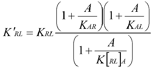
In cases where the signaling complex is a receptor-ligand complex, and the modulator is an antibody, the kinetic perturbation of the receptor-ligand interaction with an antibody can be described mathematically as:
where the change in receptor-ligand binding equilibrium constant (K'
RL) is a function of receptor-ligand equilibrium constant (K
RL), antibody concentration (A), antibody affinity for the complex (K
[RL]A) and antibody affinity for either the receptor (K
AR) or ligand (K
AL).
-
A kinetic modulator binds the target, or its signaling partner, or a complex of the target and signaling partner, in such a manner that the binding affinity or binding rate parameter of the target for its signaling partner is weakened or strengthened. For example, where the target is either a receptor or ligand, the binding affinity or binding rate parameter of the ligand for its receptor is weakened or strengthened in the presence of the kinetic modulator. A kinetic modulator with complete blocking activity represents a boundary condition in this analysis, since when K[C1C2]M is sufficiently high, K'C1C2 approaches infinity. One implication of this model is that the degree of signaling modulation is independent of kinetic modulator concentration when the concentration of kinetic modulator ([M]) is sufficiently above the equilibrium dissociation constant (KD) for the kinetic modulator/antigen interaction to be saturating for binding ligand. Hence, modulation of the interaction is related to the ratio of affinities for the complex versus the components where [M] > KD for the modulator and its antigen.
-
The present disclosure provides that the biophysical properties of a kinetic modulator's interactions with a target and/or its signaling partner can be used to predict the functional effect of the kinetic modulator on the target signaling pathway. Kinetic modulators which alter the signaling pathway can therefore be identified based on their relative affinity for target (and/or its signaling partner)in complexed versus uncomplexed form. The invention contemplates that kinetic perturbation of an interaction between two components (first component, C1 and second component, C2) of a signaling complex with a kinetic modulator (M) can be predicted in the following manner:
- K[C1C2]M or K[MC2]C1 or K[MC1]C2 < KMC2 or KMC1 leads to positive kinetic modulation
- K[C1C2]M or K[MC2]C1 or K[MC1]C2 = KMC2 or KMC1 leads to no kinetic modulation
- K[C1C2]M or K[MC2]C1 or K[MC1]C2 > KMC2 or KMC1 leads to negative kinetic modulation
-
In cases where the signaling complex is a receptor (R)-ligand(L) complex, and the kinetic modulator is an antibody (A), the kinetic perturbation can be predicted in the following manner:
- K[RL]A or K[AL]R or K[AR]L < KAL or KAR leads to positive kinetic modulation
- K[RL]A or K[AL]R or K[AR]L = KAL or KAR leads to no kinetic modulation
- K[RL]A or K[AL]R or K[AR]L > KAL or KAR leads to negative kinetic modulation
-
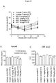
In some embodiments, a kinetic modulator, such as an antibody (A) can be identified by its ability to alter a binding interaction, such as a receptor(R)-ligand(L) interaction at any given sub-saturating concentration of the first or second component (e.g. ligand (L) concentration), as depicted in Figure 3A. The data in Figure 3A was generated from a reversible interactionmodel, assuming an affinity of the receptor ligand interactionof either 10pM, 500pM or 10nM. A kinetic modulator could effectively shift the affinity and the corresponding dose response of the receptor ligand interaction from the 500pM interaction to either the 10pM (positive modulator) or 10nM (negative modulator) as depicted. In some embodiments the kinetic modulator will produce a higher level of R-L binding at a given ligand concentration, shifting the assay curve to the left (positive modulation). In other embodiments the kinetic modulator will produce a lower level of R-L binding at a given ligand concentration, shifting the assay curve to the right (negative modulation). In some embodiments the shift is uniform, as shown in Figure 3A. In other embodiments the shift is non-uniform, reflecting the involvement of other factors e.g. accessory proteins in the complex, receptor internalization, etc. The data from Figure 3A at a 500pM affinity was used to generate Figures 3B and 3C in which the effects of various concentrations of non-agonist (Figure 3B) or agonist (Figure 3C) antibodies on signaling were depicted, assuming a fixed concentration of antigen.
-
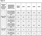
The correlation of binding characteristics to functional effect is depicted in Table 1 below for an illustrative target, insulin receptor.
Table 1 | Target Binding Characteristics | KD ratios | Functional effect |
| R | L | R-L | | |
| - | - | + | K[RL]A < KR, KL | Positive modulation |
| | | | | |
| - | + | + | K[AL]R < KL | Positive modulation |
| + | - | + | K[AL]L < KR | Positive modulation |
| | | | | |
| - | + | + | K[AL]R > KL | Negative modulation |
| + | - | + | K[AL]L > KR | Negative modulation |
-
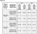
Illustrative examples of data showing the predicted effects match the binding characteristics are shown in Table 2 below.
Table 2 | Ab | Target Binding Characteristics | KD ratios | Functional effect (pAKT assay, fold-decrease in insulin EC50 relative to isotype control Ab)# |
| | R | L | R-L | | |
| Predicted | - | - | + | K[RL]A < KR, KL | Positive modulation |
| Ab078 | Out of Range* | | 3.4e-10 | | 3.3 |
| Ab085 | No Binding | | 2e-10 | | 8.9 |
| Predicted | + | - | + | K[AR]L < KR | Positive modulation |
| Ab001 | 1.2e-8 | | 1.16e-10 | 103.4 | 9.7 |
| Ab079 | 9.6e-9 | | 4.96e-10 | 19.4 | 6.7 |
| Ab080 | 1.2e-8 | | 6.8e-10 | 17.6 | 8.4 |
| Ab083 | 7.6e-9 | | 3.76e-10 | 20.2 | 8.5 |
| Predicted | + | - | + | K[AL]L = KR | Non-Modulators |
| Ab037 | 1.08e-10 | | 8e-11 | 1.4 | No change |
| Ab053 | 1.48e-10 | | 9.6e-11 | 1.5 | No change |
| Ab062 | 1.24e-10 | | 1.08e-10 | 1.1 | No change |
*Binding of this clone in the absence of insulin is evident, but insufficiently potent to be accurately measured in this assay.
#Assay run at saturating concentrations of test antibody (2-20 ug/ml). Insulin EC50 in the presence of 10 ug/ml isotype control Ab = 0.44 nM. |
-
Thus, the binding properties of the interaction(s) between the modulator and the target, its signaling partner and/or a complex comprising the target and its signaling partner, are generally predictive of the functional effect of the kinetic modulator on the target signaling pathway. Depending on the target being studied, certain other factors may need to be considered. These include: (1) the concentration of the kinetic modulator, the concentration of the target, and/or the concentration of its signaling partner (e.g., the prediction is optimized if the kinetic modulator concentration ([M]) is significantly greater than the KD of the binding between kinetic modulator and its antigen), (2) the structural form of the kinetic modulator used e.g. monovalent vs divalent or bivalent, (3) inter/intra target crosslinking, which may restrict the conformation of target and/or cause target activation, (4) the kinetic modulator's ability to alter assembly or docking, or to alter additional components of the signaling complex by steric or allosteric mechanisms, (5) signaling pathway specific properties such as alterations in the signal pathway due to disease that introduce a "bottleneck," (6) negative/positive feedback regulation of the signaling pathway, (7) alteration of clearance/internalization rates of the components of the signaling complex, (8) alterations in the target that uncouple or differentially alter ligand binding and activation e.g. a modulator enhances ligand binding but traps its receptor in a desensitized state, or a modulator attenuates ligand binding but induces a conformational change in its receptor that is activating.
-
In some aspects the invention provides methods for measuring the differential binding of a first component of a signaling complex for a second component of the signaling complex in the presence or absence of a test polypeptide agent. In these aspects, differential binding is preferably observed when there are sub-saturating concentrations of the first or second component. In some preferred embodiments the concentration of the first or second component may be reduced to provide sub-saturating conditions.
-
In some aspects the invention provides methods for measuring the differential binding of a test polypeptide binding agent, e.g. antibody, to target and/or its signaling partner, in complexed and uncomplexed form. In these aspects, differential binding is preferably observed when there are sub-saturating concentrations of test polypeptide binding agent. In some preferred embodiments the concentration of test polypeptide binding agent may be reduced to provide sub-saturating conditions.
-
In some embodiments, testing in the absence of a test polypeptide agent is performed using a control compound which is preferably a compound belonging to a similar structural class as the test polypeptide agent, but which binds to a different antigen that has no effect on the signaling complex being tested. For example, a control for a test antibody may be an isotype-matched antibody binding to an unrelated antigen, e.g keyhole limpet hemocyanin (KLH).
-
For positive modulators, at a given sub-saturating concentration of C1, higher C1 affinity will be reflected in a higher signal for C1 binding to C2 in the presence of the positive modulator. Preferential binding of the kinetic modulator will be reflected in a higher signal for the complex comprising C1 and C2, compared to the signal for either C1 alone or C2 alone. In some aspects, there may be binding of the kinetic modulator to the complex of C1 and C2, but no measurable binding to either C1 alone or C2 alone.
-
For negative modulators, at a given sub-saturating concentration of C1, lower C1 affinity will be reflected in a lower signal for C1 binding to C2 in the presence of the modulator. Preferential binding of the kinetic modulator will be reflected in a higher signal for binding of the kinetic modulator to C1 alone, or to C2 alone, compared to the signal for binding of the kinetic modulator to the complex of C1 and C2.
-
The invention provides methods of identifying a candidate polypeptide binding agent, e.g. an antibody, that modulates binding between first and second components of a signaling complex. Examples of such first and/or second components include any of the secreted proteins of Appendix A (or SEQ ID NOS: 1-88) and endogenous or exogenous signaling partners of such secreted proteins, which may be proteinaceous or non-proteinaceous but which optionally may exclude ions and salts. In some embodiments, the first and second components are polypeptides. In exemplary specific embodiments, the first and second components are endogenous.
-
Other examples include any one of TNFα, CD3, CD4, CD20, VEGF-A, CD25, HER-2, EGFR, CD33, CD52, EPO, insulin, INSR, human growth hormone, GM-CSF, G-CSF, IL-2, TPO, neurotrophic factors (NGF, NT-3, NT-4, GDNF), IFNβ, TGFβ, TNFα, FGFR4, CETP, Leptin Receptor, IL-10, IL-10 receptor alpha, IL-10 receptor beta, Growth hormone receptor, IL-13 receptor, IL-18 receptor, IL-2 receptor alpha subunit, complement factor C5a, IL-17 receptor, IL-20 receptor, IL-3 receptor, IL-4 receptor, IL-5 receptor, IL-9 receptor, Interferon type I receptor 1 (IFNAR1), Interferon type I receptor 2 (IFNAR2), Lymphocyte function antigen-3 receptor, Monocyte
chemotactic protein 1 ligand, NGF receptor, IL-6, IL-6 receptor. Their sequences are well known in the art and representative Accession Numbers and amino acid sequences from NCBI's Genbank database are set forth below. NCBI handbook [Internet]. Bethesda (MD): National Library of Medicine (US), National Center for Biotechnology Information; 2002 Oct.
Chapter 18, The Reference Sequence (RefSeq) Project. Reference to any of the proteins set forth in Appendix A or SEQ ID NOS: 1-88 herein includes reference to any naturally occurring human allelic variant thereof, such as those comprising amino acid sequences at least 90%, 91%, 92%, 93%, 94%, 95%, 96%, 97%, 98%, 99% identical to the representative sequence of any of SEQ ID NOs: 1-88, or comprising amino acid sequences encoded by nucleic acid molecules that can be obtained from human genomic DNA or cDNA libraries using nucleic acid molecules that encode any of SEQ ID NOs: 1-88 or fragments thereof that are at least about 20, 30, 40, 50 or more bases in length, e.g., under stringent hybridization conditions such as 42°C in 50% formamide, 5X SSC, 20 mM Na•PO4, pH 6.8; and washing in 1X SSC at 55°C for 30 minutes.
| Target | Accession Number | SEQ ID NO: |
| TNFα | NP_000585 | 1 |
| T cell receptor beta chain CD3 region; TCR CD3 | AAB27501 | 2 |
| CD3 antigen, delta subunit isoform B precursor | NP_001035741 | 3 |
| CD3 antigen, delta subunit isoform A precursor | NP_000723 | 4 |
| T-cell surface glycoprotein CD3 gamma chain | P09693 | 5 |
| T-cell surface glycoprotein CD3 gamma chain precursor | ACA05963 | 6 |
| T-cell surface glycoprotein CD3 epsilon chain | P07766 | 7 |
| T-cell surface glycoprotein CD3 delta chain | P04234 | 8 |
| T-cell surface glycoprotein CD3 delta chain precursor | ACA05962 | 9 |
| T-cell surface glycoprotein CD3 zeta chain | P20963 | 10 |
| CD4 | P01730 | 11 |
| CD4 antigen (p55), isoform CRA a | EAW88739 | 12 |
| CD20 | P11836 NP_068769.2 or NP 690605.1 | 13 |
| membrane-spanning 4-domains, subfamily A, member 1 | NP_690605 | 14 |
| membrane-spanning 4-domains, subfamily A, member 3 isoform a | NP_006129 | 15 |
| membrane-spanning 4-domains, subfamily A, member 3 isoform b | NP_001026979 | 16 |
| membrane-spanning 4-domains, subfamily A, member 3 isoform c | NP_001026836 | 17 |
| VEGF-A | P15692 | 18 |
| vascular endothelial growth factor A isoform a precursor | NP_001020537 | 19 |
| vascular endothelial growth factor A isoform b precursor | NP_003367 | 20 |
| vascular endothelial growth factor A isoform c precursor | NP_001020538 | 21 |
| vascular endothelial growth factor A isoform d precursor | NP_001020539 | 22 |
| vascular endothelial growth factor A isoform e precursor | NP_001020540 | 23 |
| vascular endothelial growth factor A isoform f precursor | NP_001020541 | 24 |
| vascular endothelial growth factor A isoform g precursor | NP_001028928 | 25 |
| CD25 (interleukin 2 receptor, alpha chain precursor) | NP_000408 | 26 |
| HER-2 | AAA75493 | 27 |
| EGFR | AAH94761; | 28 |
| epidermal growth factor receptor isoform a precursor | NP_005219 or P00533 | 29 |
| epidermal growth factor receptor isoform b precursor | NP_958439 | 30 |
| epidermal growth factor receptor isoform c precursor | NP 958440 | 31 |
| epidermal growth factor receptor isoform d precursor | NP_958441 | 32 |
| CD33 antigen isoform 1 precursor | NP_001763 | 33 |
| CD33 antigen isoform 2 precursor | NP_001076087 | 34 |
| CD33 antigen (gp67), isoform CRA a | EAW71994 | 35 |
| CD33 antigen (gp67), isoform CRA b | EAW71995 | 36 |
| CD33 antigen (gp67), isoform CRA c | EAW71996 | 37 |
| CD52 antigen precursor | NP_001794 | 38 |
| EPO | CAA26095 | 39 |
| insulin | AAA59172 | 40 |
| INSR | P06213 | 41 |
| insulin receptor isoform Short precursor | NP_001073285 | 42 |
| insulin receptor isoform Long precursor | NP_000199 | 43 |
| human growth hormone | AAA72260 | 44 |
| GM-CSF | AAA52578 | 45 |
| G-CSF | P09919 | 46 |
| IL-2 | AAB46883 | 47 |
| TPO | AAB33390 | 48 |
| NGF | AAH32517 | 49 |
| nerve growth factor, beta polypeptide precursor | NP_002497 | 50 |
| NT-3 | P20783 | 51 |
| neurotrophin 3 isoform 1 preproprotein | NP_001096124 | 52 |
| neurotrophin 3 isoform 2 preproprotein | NP_002518 | 53 |
| NT-4 | AAA60154 | 54 |
| Chain A, Brain Derived Neurotrophic Factor, Neurotrophin-4 | 1B8M_A | 55 |
| Chain B, Brain Derived Neurotrophic Factor, Neurotrophin-4 | 1B8M_B | 56 |
| GDNF | P39905 | 57 |
| glial cell derived neurotrophic factor isoform 1 preproprotein | NP_000505 | 58 |
| glial cell derived neurotrophic factor isoform 2 precursor | NP_954701 | 59 |
| glial cell derived neurotrophic factor isoform 3 | NP_954704 | 60 |
| IFNβ | P01574 | 61 |
| TGFβ | AAA36738 | 62 |
| FGFR4 | AAB25788 | 63 |
| fibroblast growth factor receptor 4 isoform 1 precursor | NP_998812 | 64 |
| fibroblast growth factor receptor 4 isoform 2 precursor | NP_075252 | 65 |
| CETP | P11597 | 66 |
| Leptin Receptor | P48357 | 67 |
| IL-10 | P22301 | 68 |
| IL-10 receptor alpha | EAW67343 | 69 |
| IL-10 receptor beta | AAH01903 | 70 |
| Growth hormone receptor | P10912 | 71 |
| IL-13 receptor | CAA70021 | 72 |
| IL-18 receptor | AAH93977 | 73 |
| IL-2 receptor alpha subunit | P01589 | 74 |
| complement factor C5a | NP_001726 | 75 |
| IL-17 receptor | AAB99730 | 76 |
| IL-20 receptor | Q9UHF4 | 77 |
| IL-3 receptor | AAA59148 | 78 |
| IL-4 receptor | CAA36672 | 79 |
| IL-5 receptor | CAA01794 | 80 |
| IL-9 receptor | AAB30844 | 81 |
| Interferon type I receptor 1 (IFNAR1) | P17181 | 82 |
| Interferon type I receptor 2 (IFNAR2) | P48551 | 83 |
| Lymphocyte function antigen-3 receptor | P19256 | 84 |
| Monocyte chemotactic protein 1 ligand | P13500 or | 85 |
| | NP_002973 | |
| NGF receptor | AAB59544 | 86 |
| IL-6 | NP_000591 | 87 |
| IL-6 receptor | NP_000556 | 88 |
-
In one aspect, the methods of identifying a candidate kinetic modulating drug include (a) measuring a binding affinity or binding rate parameter of said first component for said second component, in the presence of a test polypeptide binding agent, e.g. antibody, (b) measuring a binding affinity or binding rate parameter of said first component for said second component in the absence of said test polypeptide binding agent; and (c) identifying said test polypeptide binding agent as a candidate kinetic modulating drug when said test polypeptide binding agent exhibits at least a 1.5-fold difference in the binding affinity or binding rate parameter measured in steps (a) and (b). In some embodiments, the difference in binding affinity or binding rate parameter ranges from about 1.5-fold (i.e., 50%) to, optionally, about 1000-fold, or about 1.5-fold to about 100-fold, or about 2-fold to 25-fold, or about 2-fold to about 50-fold, or about 1.5-fold to about 25-fold, or about 1.5-fold to about 50-fold.
-
In some embodiments, the test polypeptide binding agent is identified as a candidate positive modulator if the test polypeptide agent strengthens the binding affinity or binding rate parameter between said first component and said second component. In other embodiments, the test polypeptide agent is identified as a candidate negative modulator if the test polypeptide agent weakens the binding affinity or binding rate parameter between said first component and said second component.
-
Whether a change (increase or decrease) in a particular binding affinity value or binding rate parameter value represents "strengthened" (or stronger) or "weakened" (or weaker) binding affinity or binding rate parameter depends on the value of the parameter and its units, and is well known in the art. For example, in the case of the parameter KA, higher values mean "strengthened" binding affinity, such that a KA of about 106M-1 is stronger than a KA of about 105M-1. As another example, in the case of the parameter KD, lower values mean "strengthened" binding affinity, such that a KD of about 10-6M is stronger than a KD of about 10-5M. Conversely, in the case of KA, lower values mean "weakened" binding affinity, such that a KA of about 105M-1 is a weakened binding affinity compared to a KA of about 106M-1. As another example, in the case of KD, higher values mean "weakened" binding affinity, such that a KD of about 10-5M is weakened binding affinity compared to a KD of about 10-6M.
-
As used herein, a "strengthened" binding rate parameter means increased residency time, faster association or slower dissociation. As used herein, a "weakened" binding rate parameter means decreased residency time, slower association or faster dissociation.
-
Binding affinity can also be determined through the ratio of the on-rate and off-rate binding rate parameters. Generally, in the case of on-rate, higher values mean faster or stronger association or increased residence time, and typically result in stronger binding affinity. Conversely, lower values for on-rate mean slower or weaker association or decreased residence time, and typically result in weaker binding affinity. Generally, in the case of off-rate, higher values mean faster dissociation or decreased residence time, and typically result in weaker binding affinity. Conversely, lower values for off-rate mean slower dissociation or increased residence time, and typically result in stronger binding affinity. This is because the ratio of off-rate to on-rate, or on-rate to off-rate, indicates binding affinity as displayed in the equations below.
where
-
Even when binding affinity is not detectably or significantly altered, however, the change in residence time, i.e. an increased residence time via increased on-rate or decreased off-rate, or a decreased residence time via a decreased on-rate or increased off-rate, may still result in differential activation of signaling pathways. For example, in some instances where a receptor may activate two different pathways, the pathways differ in the degree of receptor activation required for a full effect. One signaling pathway can be fully activated at low levels of receptor activation or residence time, while full activation of the second pathway requires higher levels of receptor activation or residence time.
-
In another aspect, the methods of identifying a candidate kinetic modulating drug include (a) (i) measuring a binding affinity or binding rate parameter of a test polypeptide binding agent, e.g. antibody, for said first component in the presence of said second component, or (ii) measuring a binding affinity or binding rate parameter of a test polypeptide binding agent for said second component in the presence of said first component; and (b) (i) measuring a binding affinity or binding rate parameter of said test polypeptide binding agent for said first component in the absence of said second component, or (ii) measuring a binding affinity or binding rate parameter of said test polypeptide binding agent for said second component in the absence of said first component; and (c) identifying said test polypeptide binding agent as a candidate kinetic modulating drug when said test polypeptide binding agent exhibits at least a 1.5-fold (i.e., 50%) difference in the binding affinity or binding rate parameters measured in steps (a) and (b).
-
In some embodiments, the test polypeptide binding agent is identified as a candidate positive modulator if the binding affinity or binding rate parameter measured in step (a) is at least 1.5-fold (i.e., 50%) stronger than the binding affinity or binding rate parameter measured in step (b). In specific embodiments, the binding affinity or binding rate parameter measured in step (a) compared to that measured in step (b) is about 1.5-fold (i.e., 50%) to, optionally, about 1000-fold stronger for step (a) vs. step (b), or about 1.5-fold to about 100-fold, or about 2-fold to 25-fold, or about 2-fold to about 50-fold, or about 1.5-fold to about 25-fold, or about 1.5-fold to about 50-fold, e.g. at least 1.5-fold, 2-fold, 3-fold, 4-fold, 5-fold, 6-fold, 7-fold, 8-fold, 9-fold, 10-fold, 11-fold, 12-fold, 13-fold, 14-fold, 15-fold, 16-fold, 17-fold, 18-fold, 19-fold or 20-fold, or up to 500-fold, or up to 200-fold, or up to 150-fold, or up to 100-fold, or up to 90-fold, or up to 80-fold, or up to 70-fold, or up to 60-fold, or up to 50-fold, or up to 40-fold, or up to 30-fold.
-
In other embodiments, the test polypeptide binding agent is identified as a candidate negative modulator if the binding affinity or binding rate parameter measured in step (b) is at least 1.5-fold (i.e., 50%) stronger than the binding affinity or binding rate parameter measured in step (a). In specific embodiments, the binding affinity or binding rate parameter measured in step (b) compared to that measured in step (a) is about 1.5-fold (i.e., 50%) to, optionally, about 1000-fold stronger for step (b) vs. step (a), or about 1.5-fold to about 100-fold, or about 2-fold to 25-fold, or about 2-fold to about 50-fold, or about 1.5-fold to about 25-fold, or about 1.5-fold to about 50-fold, e.g. at least 1.5-fold, 2-fold, 3-fold, 4-fold, 5-fold, 6-fold, 7-fold, 8-fold, 9-fold, 10-fold, 11-fold, 12-fold, 13-fold, 14-fold, 15-fold, 16-fold, 17-fold, 18-fold, 19-fold or 20-fold, or up to 500-fold, or up to 200-fold, or up to 150-fold, or up to 100-fold, or up to 90-fold, or up to 80-fold, or up to 70-fold, or up to 60-fold, or up to 50-fold, or up to 40-fold, or up to 30-fold.
-
In some embodiments, the binding affinity or binding rate parameter of the test polypeptide binding agent for the first component alone is measured. In some embodiments, the binding affinity or binding rate parameter of the test polypeptide binding agent for the second component alone is measured.
-
In some embodiments, the test polypeptide binding agent is identified as a candidate positive modulator if one or more binding affinity or binding rate parameters selected from the group consisting of (A) the binding affinity or binding rate parameter of the test polypeptide binding agent for a complex comprising the first and second components, optionally K[C1C2]M, (B) the binding affinity or binding rate parameter of the first component for a complex comprising the polypeptide binding agent and the second component, optionally K[MC2]C1, or (C) the binding affinity or binding rate parameter of the second component for a complex comprising the polypeptide binding agent and the first component, optionally K[MC1]C2, is at least about 1.5-fold stronger than one or more binding affinity or binding rate parameter selected from the group consisting of (1) the binding affinity or binding rate parameter of the test polypeptide binding agent for the second component alone, optionally KMC2 or (2) the binding affinity or binding rate parameter of the test polypeptide binding agent for the first component alone, optionally KMC1. In some embodiments, the specific binding affinity or binding rate parameter of any one or more of (A), (B) or (C) is about 1.5-fold (i.e., 50%) to, optionally, about 1000-fold stronger than the binding affinity or binding rate parameter of any one or more of (1) or (2); or alternatively, about 1.5-fold to about 100-fold stronger, or about 2-fold to 25-fold, or about 2-fold to about 50-fold, or about 1.5-fold to about 25-fold, or about 1.5-fold to about 50-fold, e.g. at least 1.5-fold, 2-fold, 3-fold, 4-fold, 5-fold, 6-fold, 7-fold, 8-fold, 9-fold, 10-fold, 11-fold, 12-fold, 13-fold, 14-fold, 15-fold, 16-fold, 17-fold, 18-fold, 19-fold or 20-fold, or up to 500-fold, or up to 200-fold, or up to 150-fold, or up to 100-fold, or up to 90-fold, or up to 80-fold, or up to 70-fold, or up to 60-fold, or up to 50-fold, or up to 40-fold, or up to 30-fold. For example, in some embodiments, the binding affinity or binding rate parameter of any one or more of (A), (B) or (C) is stronger than the binding affinity or binding rate parameter of both (1) and (2). In some embodiments, the binding affinity or binding rate parameter of (1) is stronger than the binding affinity or binding rate parameter of (2). In other embodiments, the binding affinity or binding rate parameter of (2) is stronger than the binding affinity or binding rate parameter of (1). In some embodiments, two or more binding affinity or binding rate parameters are measured and compared, e.g. off-rate and on-rate, or KA and KD, or any combination thereof.
-
In specific embodiments, wherein the binding affinity measured is the equilibrium dissociation constant KD, any of K[C1C2]M, K[MC2]C1, or K[MC1]C2 is lower, e.g., about 1.5-fold to, optionally, 1000-fold lower, than any of KMC2 or KMC1. Similarly, wherein the binding affinity measured is the off-rate, any of the off-rates between (A) [C1C2] and M, or (B) [MC2] and C1, or (C) [MC1] and C2 are lower, e.g. about 1.5-fold to, optionally, 1000-fold lower, than any of the off-rates between (1) M and C2 or (2) M and C1. In one exemplary embodiment, K[C1C2]M is about 1.5-fold to, optionally, 1000-fold lower than KMC2. In another exemplary embodiment, K[MC2]C1 is about 1.5-fold to, optionally, 1000-fold lower than KMC2. In another exemplary embodiment, K[MC1]C2 is about 1.5-fold to, optionally, 1000-fold lower than KMC2. In another exemplary embodiment, K[C1C2]M is about 1.5-fold to, optionally, 1000-fold lower than KMC1. In another exemplary embodiment, K[MC2]C1 is about 1.5-fold to, optionally, 1000-fold lower than KMC1. In yet another exemplary embodiment, K[MC1]C2 is about 1.5-fold to, optionally, 1000-fold lower than KMC1. Similar examples can be envisioned for each of the off-rates between (A) [C1C2] and M, or (B) [MC2] and C1, or (C) [MC1] and C2, compared to each of the off-rates between (1) M and C2 or (2) M and C1.
-
Conversely, where the binding affinity measured is the equilibrium association constant KA, any of K[C1C2]M, K[MC2]C1, or K[MC1]C2 is higher, e.g., about 1.5-fold to, optionally, 1000-fold higher, than any of KMC2 or KMC1. Similarly, wherein the binding affinity measured is the on-rate, any of the on-rates between (A) [C1C2] and M, or (B) [MC2] and C1, or (C) [MC1] and C2 are higher, e.g. about 1.5-fold to, optionally, 1000-fold higher, than any of the on-rates between (1) M and C2 or (2) M and C1. In one exemplary embodiment, K[C1C2]M is about 1.5-fold to, optionally, 1000-fold higher than KMC2. In another exemplary embodiment, K[MC2]C1 is about 1.5-fold to, optionally, 1000-fold higher than KMC2. In another exemplary embodiment, K[MC1]C2 is about 1.5-fold to, optionally, 1000-fold higher than KMC2. In another exemplary embodiment, K[C1C2]M is about 1.5-fold to, optionally, 1000-fold higher than KMC1. In another exemplary embodiment, K[MC2]C1 is about 1.5-fold to, optionally, 1000-fold higher than KMC1. In yet another exemplary embodiment, K[MC1]C2 is about 1.5-fold to, optionally, 1000-fold higher than KMC1. Similar examples can be envisioned for each of the on-rates between (A) [C1C2] and M, or (B) [MC2] and C1, or (C) [MC1] and C2, compared to each of the on-rates between (1) M and C2 or (2) M and C1.
-
In some embodiments, the test polypeptide binding agent is identified as a candidate negative modulator if one or more binding affinity or binding rate parameters selected from the group consisting of (1) the binding affinity or binding rate parameter of the test polypeptide binding agent for the second component alone, optionally KMC2, or (2) the binding affinity or binding rate parameter of the test polypeptide binding agent for the first component alone, optionally KMC1, is at least about 1.5-fold stronger than one or more binding affinity or binding rate parameter selected from the group consisting of (A) the binding affinity or binding rate parameter of the test polypeptide binding agent for a complex comprising the first and second components, optionally K[C1C2]M, (B) the binding affinity or binding rate parameter of the first component for a complex comprising the polypeptide binding agent and the second component, optionally K[MC2]C1, or (C) the binding affinity or binding rate parameter of the second component for a complex comprising the polypeptide binding agent and the first component, optionally K[MC1]C2. In some embodiments, the specific binding affinity or binding rate parameter of any one or more of (1) or (2) is about 1.5-fold (i.e., 50%) to, optionally, about 1000-fold stronger than the binding affinity or binding rate parameter of any one or more of (A), (B) or (C); or alternatively, about 1.5-fold to about 100-fold stronger, or about 2-fold to 25-fold, or about 2-fold to about 50-fold, or about 1.5-fold to about 25-fold, or about 1.5-fold to about 50-fold, e.g. at least 1.5-fold, 2-fold, 3-fold, 4-fold, 5-fold, 6-fold, 7-fold, 8-fold, 9-fold, 10-fold, 11-fold, 12-fold, 13-fold, 14-fold, 15-fold, 16-fold, 17-fold, 18-fold, 19-fold or 20-fold, or up to 500-fold, or up to 200-fold, or up to 150-fold, or up to 100-fold, or up to 90-fold, or up to 80-fold, or up to 70-fold, or up to 60-fold, or up to 50-fold, or up to 40-fold, or up to 30-fold. In some embodiments, the binding affinity or binding rate parameter of any of (1) or (2) is stronger than the binding affinity or binding rate parameter of all of (A), (B) and (C). In some embodiments, the binding affinity or binding rate parameter of (1) is stronger than the binding affinity or binding rate parameter of (2). In other embodiments, the binding affinity or binding rate parameter of (2) is stronger than the binding affinity or binding rate parameter of (1). In some embodiments, two or more binding affinity or binding rate parameters are measured and compared, e.g. off-rate and on-rate, or KA and KD, or any combination thereof.
-
In specific embodiments, where the binding affinity measured is the equilibrium dissociation constant KD, any of KMC2 or KMC1 is lower, e.g., about 1.5-fold to, optionally, 1000-fold lower, than any of K[C1C2]M, K[MC2]C1, or K[MC1]C2. Similarly, wherein the binding affinity measured is the off-rate, any of the off-rates between (1) M and C2 or (2) M and C1 are lower, e.g. about 1.5-fold to, optionally, 1000-fold lower, than any of the off-rates between (A) [C1C2] and M, or (B) [MC2] and C1, or (C) [MC1] and C2. In one exemplary embodiment KMC2 is about 1.5-fold to, optionally, 1000-fold lower than K[C1C2]M. In another exemplary embodiment, KMC2 is about 1.5-fold to, optionally, 1000-fold lower than K[MC2]C1. In another exemplary embodiment, KMC2 is about 1.5-fold to, optionally, 1000-fold lower than K[MC1]C2. In another exemplary embodiment, KMC1 is about 1.5-fold to, optionally, 1000-fold lower than K[C1C2]M. In another exemplary embodiment, KMC1 is about 1.5-fold to, optionally, 1000-fold lower than K[MC2]C1. In yet another exemplary embodiment, KMC1 is about 1.5-fold to, optionally, 1000-fold lower than K[MC1]C2. Similar examples can be envisioned for each of the off-rates between (1) M and C2 or (2) M and C1, compared to each of the off-rates between (A) [C1C2] and M, or (B) [MC2] and C1, or (C) [MC1] and C2.
-
Conversely, wherein the binding affinity is the equilibrium association constant KA, any of KMC2 or KMC1 is higher, e.g., about 1.5-fold to, optionally, 1000-fold higher, than any of K[C1C2]M, K[MC2]C1, or K[MC1]C2. Similarly, wherein the binding affinity measured is the on-rate, any of the on-rates between (1) M and C2 or (2) M and C1 are higher, e.g. about 1.5-fold to, optionally, 1000-fold higher, than any of the on-rates between (A) [C1C2] and M, or (B) [MC2] and C1, or (C) [MC1] and C2. In one exemplary embodiment KMC2 is about 1.5-fold to, optionally, 1000-fold higher than K[C1C2]M. In another exemplary embodiment, KMC2 is about 1.5-fold to, optionally, 1000-fold higher than K[MC2]C1. In another exemplary embodiment, KMC2 is about 1.5-fold to, optionally, 1000-fold higher than K[MC1]C2. In another exemplary embodiment, KMC1 is about 1.5-fold to, optionally, 1000-fold higher than K[C1C2]M. In another exemplary embodiment, KMC1 is about 1.5-fold to, optionally, 1000-fold higher than K[MC2]C1. In yet another exemplary embodiment, KMC1 is about 1.5-fold to, optionally, 1000-fold higher than K[MC1]C2. Similar examples can be envisioned for each of the on-rates between (1) M and C2 or (2) M and C1, compared to each of the on-rates between (A) [C1C2] and M, or (B) [MC2] and C1, or (C) [MC1] and C2.
-
In any of these embodiments, the test polypeptide binding agent and second component can be contacted with multiple different concentrations of said first component. In any of these embodiments, the test polypeptide binding agent and first component can be contacted with multiple different concentrations of said second component. In any of these embodiments, multiple different concentrations of the test polypeptide binding agent can be contacted with said first component and said second component.
-
When the effect of test polypeptide binding agent on the binding interaction between the first component and second component is determined, in some specific embodiments, when the antigen for the test polypeptide binding agent is the first component, e.g., ligand, the test polypeptide binding agent is at a saturating concentration compared to the concentration of the first component. Alternatively, when the antigen for the test polypeptide binding agent is the second component, e.g., receptor, the test polypeptide binding agent is at a saturating concentration compared to the concentration of the second component. In some embodiments, the concentration of the test polypeptide binding agent is greater than or equal to the KD of the test polypeptide binding agent for a complex comprising the first component and the second component. In further embodiments, the concentration of the second component is less than the KD of the test polypeptide binding agent for the first component, e.g., ligand. In yet further embodiments, the concentration of the first component, e.g., ligand, is at a subsaturating concentration for the binding of first component to second component, e.g., receptor. In some embodiments, the concentration of the first component, e.g., ligand is within the range of about EC20 to EC80 for the interaction of the first component with the second component. In some embodiments, one or more concentrations of the test polypeptide binding agent is contacted with multiple different concentrations of the first component, e.g., ligand, in the presence of one or more concentrations of the second component, e.g., receptor. In some embodiments, one or more concentrations of the test polypeptide binding agent is contacted with multiple different concentrations of the second component, e.g., receptor, in the presence of one or more concentrations of the first component, e.g., ligand.
-
When differential binding of test polypeptide binding agent to complexed vs uncomplexed target and/or signaling partner is determined in order to identify a positive modulator, in some embodiments, the test polypeptide binding agent is at a saturating concentration for a complex comprising the first component and the second component. In some embodiments, the concentration of test polypeptide binding agent is greater than or equal to the KD of the test polypeptide binding agent for a complex comprising the first component, e.g., ligand, and the second component, e.g., receptor. In further embodiments, the concentration of the second component, e.g., receptor is greater than the KD of the second component, e.g., receptor, for the first component, e.g., ligand. In further embodiments, the concentration of the first component, e.g., ligand, is a saturating concentration for the second component, e.g., receptor. In yet further embodiments, the test polypeptide binding agent is at a subsaturating concentration for a complex comprising the first component and the second component. In some embodiments, the concentration of the polypeptide binding agent is within the range of about EC20 to EC80 for the interaction of the first component with the second component. In some embodiments, the concentration of the second component, e.g., receptor, is greater than the KD of the second component, e.g., receptor, for the first component, e.g., ligand. In some embodiments, the concentration of the first component, e.g., ligand, is a saturating concentration for the second component, e.g., receptor.
-
When differential binding of test polypeptide binding agent to complexed vs uncomplexed target and/or signaling partner is determined in order to identify a negative modulator, in some embodiments, when the antigen to which the test polypeptide binding agent binds is the first component, e.g., ligand, the test polypeptide binding agent is at a subsaturating concentration for the first component. When the antigen to which the test polypeptide binding agent binds is the second component, e.g., receptor, the test polypeptide binding agent is at a subsaturating concentration for the second component. In further embodiments, the concentration of the polypeptide binding agent is within the range of about EC20 to EC80 for the interaction of the first component with the second component. In further embodiments, the concentration of the second component, e.g., receptor, is greater than the KD of the second component, e.g., receptor, for the first component, e.g., ligand. In further embodiments, the concentration of the first component, e.g., ligand, is a saturating concentration for the second component, e.g., receptor.
-
In some embodiments, the methods further involve assaying a plurality of test polypeptide binding agents, e.g. antibodies, for binding affinity to any one of (a) the first component, (b) the second component, or (c) a complex comprising the first component and second component. In some specific embodiments, the polypeptide binding agents have a binding affinity characterized, e.g., by an equilibrium dissociation constant KD of about 10-5M or less, or about 10-6M or less, or about 10-7M or less, or about 10-8M or less, where a lower KD means stronger binding affinity. In some embodiments, the plurality of test polypeptide binding agents screened are variants of a parent polypeptide binding agent made by introducing one or more different mutations into a parent polypeptide binding agent.
-
In further embodiments, the polypeptide binding agents may be screened for selectivity of effect for the first or second component, compared to a different binding partner such as a decoy receptor, clearance receptor, or alternate signal pathway component. Such methods may involve identifying a polypeptide binding agent that does not significantly change the binding affinity or binding rate parameter of the first or second component for a different binding partner, such binding partner being neither the first nor second component. In some embodiments, the presence of the polypeptide binding agent changes the binding affinity or binding rate parameter of the first or second component for a different binding partner no more than 5-fold, or no more than 10-fold, or no more than 20-fold, or no more than 30-fold, or no more than 40-fold, or no more than 50-fold.
-
Any of the preceding methods may further include measuring the level of signaling mediated by the signaling complex in the presence and absence of the test polypeptide binding agent, and determining whether the test polypeptide binding agent is additionally an agonist, partial agonist, antagonist or partial antagonist. Antagonism or agonism can be measured in any in vitro or in vivo assay known in the art, including but not limited to signaling in a phosphorylation assay, ion flux assay, molecular transport assay, or gene expression assay.
-
In some embodiments, the test polypeptide binding agent shifts (positively or negatively) the dose-response curve of the interaction of the first component, e.g. ligand, with the second component, e.g. receptor. The shift may manifest as an increased or decreased EC50 by at least about 1.5-fold, e.g. about 1.5-fold to about 1000-fold. In some embodiments, the test polypeptide binding agent does not significantly change the maximal agonist response of the signal produced by interaction of the first and second components of the signaling complex. In other embodiments, the test polypeptide binding agent itself acts as an antagonist (e.g., reduces the maximal agonist response of the signaling produced by said signaling complex) or agonist (e.g. increases the maximal agonist response of the signaling produced by said signaling complex).
-
Where the test polypeptide binding agent acts as an antagonist or partial antagonist, the maximal agonist response may be decreased, e.g., by about 1.5-fold to about 100-fold, or about 2-fold to about 25-fold, or about 1.5-fold to about 50-fold; or, decreased by about 10%, 25%, 50% (1.5-fold), 75%, 2-fold, 3-fold, or 4-, 5-, 6-, 7-, 8-, 9- or 10-fold. Alternatively, where the test polypeptide binding agent acts as an agonist or partial agonist, the maximal agonist response may be increased, e.g. by at least about 10%, 25%, 50% (1.5-fold), 75%, 2-fold, 3-fold, or 4-, 5-, 6-, 7-, 8-, 9- or 10-fold. Moreover, when the test polypeptide binding agent acts as an antagonist or partial antagonist, the IC50 may be 1x10⁻⁵ or less. The test polypeptide binding agent may exhibit further desirable characteristics, e.g. the test polypeptide binding agent does not significantly decrease clearance of said first component, or said second component, or said signaling complex comprising said first and second components.
-
In a related aspect, the invention provides an antibody identified by any of the methods described above or anywhere in the present application.
Polypeptide bending agents with desired characteristics
-
The invention also provides polypeptide binding agents, e.g., antibodies, that possess certain desirable characteristics. In some embodiments, the invention provides a positive modulator that (a) binds to the target, e.g., the secreted protein of any of Appendix A (or SEQ ID NOS: 1-88) or any of the ligands, receptors or components described herein, with an equilibrium dissociation constant KD of about 10-5M or less, e.g., 10-6M or less, or 10-7M or less, or 10-8M or less, and (b) is capable of improving the binding affinity KD between said target and its signaling partner by at least about 1.5-fold (i.e., 50%); or by about 1.5-fold to, optionally, about 1000-fold, or 1.5-fold to about 100-fold, or about 2-fold to 25-fold, or about 2-fold to about 50-fold, or about 1.5-fold to about 25-fold, or about 1.5-fold to about 50-fold, e.g. at least about 1.5-fold, 2-fold, 3-fold, 4-fold, 5-fold, 6-fold, 7-fold, 8-fold, 9-fold, 10-fold, 11-fold, 12-fold, 13-fold, 14-fold, 15-fold, 16-fold, 17-fold, 18-fold, 19-fold or 20-fold, or up to about 500-fold, or up to about 200-fold, or up to about 150-fold, or up to about 100-fold, or up to about 90-fold, or up to about 80-fold, or up to about 70-fold, or up to about 60-fold, or up to about 50-fold, or up to about 40-fold, or up to about 30-fold.
-
In other embodiments, the invention provides a negative modulator that (a) binds to the target, e.g. the secreted protein of any of Appendix A (or SEQ ID NOS: 1-88) or any of the ligands, receptors or components described herein, with an equilibrium dissociation constant KD of about 10-5M or less, e.g., 10-6M or less, or 10-7M or less, or 10-8M or less, and (b) is capable of reducing the binding affinity KD between said secreted protein and its signaling partner by at least about 1.5-fold (i.e., 50%); or by about 1.5-fold to, optionally, about 1000-fold, or 1.5-fold to about 100-fold, or about 2-fold to 25-fold, or about 2-fold to about 50-fold, or about 1.5-fold to about 25-fold, or about 1.5-fold to about 50-fold, e.g. at least about 1.5-fold, 2-fold, 3-fold, 4-fold, 5-fold, 6-fold, 7-fold, 8-fold, 9-fold, 10-fold, 11-fold, 12-fold, 13-fold, 14-fold, 15-fold, 16-fold, 17-fold, 18-fold, 19-fold or 20-fold, or up to about 500-fold, or up to about 200-fold, or up to about 150-fold, or up to about 100-fold, or up to about 90-fold, or up to about 80-fold, or up to about 70-fold, or up to about 60-fold, or up to about 50-fold, or up to about 40-fold, or up to about 30-fold.
-
In some embodiments, the invention provides a positive modulating antibody that strengthens the binding of a first component (C1) to a second component (C2) of a signaling complex, said antibody characterized by the following equilibrium dissociation constant KD binding properties: (i) said antibody binds with an equilibrium dissociation constant KD of about 10-5M or less, e.g., 10-6M or less, or 10-7M or less, or 10-8M or less, to any one of C1, C2, or a complex comprising C1 and C2 (C1C2); and (ii) any of K[C1C2]A, K[AC2]C1, or K[AC1]C2 is at least about 50% (1.5-fold) lower than any of KAC2 or KAC1, wherein C1 or C2 is a target and its signaling partner, optionally the secreted protein of any of Appendix A(or SEQ ID NOS: 1-88). In some embodiments any of K[C1C2]A, K[AC2]C1, or K[AC1]C2 is about 1.5-fold to, optionally, about 1000-fold lower than any of KAC2 or KAC1; or 1.5-fold to about 100-fold lower, or about 2-fold to 25-fold, or about 2-fold to about 50-fold, or about 1.5-fold to about 25-fold, or about 1.5-fold to about 50-fold, e.g. at least about 1.5-fold, 2-fold, 3-fold, 4-fold, 5-fold, 6-fold, 7-fold, 8-fold, 9-fold, 10-fold, 11-fold, 12-fold, 13-fold, 14-fold, 15-fold, 16-fold, 17-fold, 18-fold, 19-fold or 20-fold, or up to about 500-fold, or up to about 200-fold, or up to about 150-fold, or up to about 100-fold, or up to about 90-fold, or up to about 80-fold, or up to about 70-fold, or up to about 60-fold, or up to about 50-fold, or up to about 40-fold, or up to about 30-fold lower. In some embodiments, any of K[C1C2]A, K[AC2]C1, or K[AC1]C2 is at least about 1.5-fold lower than both of KAC2 or KAC1; or 1.5-fold to, optionally, about 1000-fold lower, or 1.5-fold to about 100-fold lower, or about 2-fold to 25-fold, or about 2-fold to about 50-fold, or about 1.5-fold to about 25-fold, or about 1.5-fold to about 50-fold, e.g. at least about 1.5-fold, 2-fold, 3-fold, 4-fold, 5-fold, 6-fold, 7-fold, 8-fold, 9-fold, 10-fold, 11-fold, 12-fold, 13-fold, 14-fold, 15-fold, 16-fold, 17-fold, 18-fold, 19-fold or 20-fold, or up to about 500-fold, or up to about 200-fold, or up to about 150-fold, or up to about 100-fold, or up to about 90-fold, or up to about 80-fold, or up to about 70-fold, or up to about 60-fold, or up to about 50-fold, or up to about 40-fold, or up to about 30-fold lower.
-
In some embodiments, the invention provides a negative modulating antibody that weakens the binding of a first component (C1) to a second component (C2) of a signaling complex, said antibody characterized by the following equilibrium dissociation constant KD binding properties: (i) said antibody binds with an equilibrium dissociation constant KD of about 10-5M or less, e.g., 10-6M or less, or 10-7M or less, or 10-8M or less, to any one of C1, C2, or a complex comprising C1 and C2 (C1C2), and (ii) any of KAC2 or KAC1 is at least about 50% (1.5-fold) lower than any of K[C1C2]A, K[AC2]C1, or K[AC1]C2, wherein C1 or C2 is a target and its signaling partner, optionally the secreted protein of any of Appendix A (or SEQ ID NOS: 1-88). In some embodiments, any of KAC2 or KAC1 is at least about 1.5-fold to, optionally, 1000-fold lower than any of K[C1C2]A, K[AC2]C1, or K[AC1]C2; or 1.5-fold to about 100-fold lower, or about 2-fold to 25-fold, or about 2-fold to about 50-fold, or about 1.5-fold to about 25-fold, or about 1.5-fold to about 50-fold, e.g. at least about 1.5-fold, 2-fold, 3-fold, 4-fold, 5-fold, 6-fold, 7-fold, 8-fold, 9-fold, 10-fold, 11-fold, 12-fold, 13-fold, 14-fold, 15-fold, 16-fold, 17-fold, 18-fold, 19-fold or 20-fold, or up to about 500-fold, or up to about 200-fold, or up to about 150-fold, or up to about 100-fold, or up to about 90-fold, or up to about 80-fold, or up to about 70-fold, or up to about 60-fold, or up to about 50-fold, or up to about 40-fold, or up to about 30-fold lower. In some embodiments, any of KAC2 or KAC1 is at least about 1.5-fold lower than all of K[C1C2]A, K[AC2]C1, or K[AC1]C2; or 1.5-fold to, optionally, about 1000-fold lower, or 1.5-fold to about 100-fold lower, or about 2-fold to 25-fold, or about 2-fold to about 50-fold, or about 1.5-fold to about 25-fold, or about 1.5-fold to about 50-fold, e.g. at least about 1.5-fold, 2-fold, 3-fold, 4-fold, 5-fold, 6-fold, 7-fold, 8-fold, 9-fold, 10-fold, 11-fold, 12-fold, 13-fold, 14-fold, 15-fold, 16-fold, 17-fold, 18-fold, 19-fold or 20-fold, or up to about 500-fold, or up to about 200-fold, or up to about 150-fold, or up to about 100-fold, or up to about 90-fold, or up to about 80-fold, or up to about 70-fold, or up to about 60-fold, or up to about 50-fold, or up to about 40-fold, or up to about 30-fold lower.
-
Any of such polypeptide binding agents are preferably purified and substantially homogeneous, e.g. at least about 90%, 95%, 97%, 98%, 99% or 99.5% pure. In some examples, the polypeptide binding agents are monoclonal antibodies.
-
The invention further provides methods of preparing a sterile pharmaceutical composition comprising adding a sterile pharmaceutically acceptable diluent to such polypeptide binding agents, sterile compositions of such polypeptide binding agents, e.g., in a therapeutically effective amount, and methods of administering such sterile compositions, e.g. to modulate (increase or decrease) signaling of a complex comprising the secreted protein.
Signaling complexes
-
Activation of genes, alterations in metabolism, the continued proliferation and death of the cell, and the stimulation or suppression of locomotion, are some of the cellular responses to extracellular stimulation that may be mediated by signaling complexes. Gene activation leads to further cellular effects, since the protein products of many of the responding genes include enzymes and transcription factors themselves. Transcription factors produced as a result of a signal transduction cascade can, in turn, activate yet more genes. Therefore an initial stimulus can trigger the expression of an entire cohort of genes, and this, in turn, can lead to the activation of any number of complex physiological events. These events include the increased uptake of glucose from the blood stream stimulated by insulin and the migration of neutrophils to sites of infection stimulated by bacterial products.
-
Neurotransmitters are ligands that are capable of binding to ion channel proteins to form a complex, resulting in their opening to allow the rapid flow of a particular ion across the plasma membrane. This results in an altering of the cell's membrane potential and is important for processes such as the neural conduction of electrochemical impulses. For example, binding of the neurotransmitter acetylcholine at certain synapses opens channels that admit Na+ and initiate a nerve impulse or muscle contraction. Ligands can be freely soluble, or can be found on the surface of other cells or within the extracellular matrix. Such cell surface or extracellular matrix ligands signal between cells when they come in contact with each other, such as when a phagocytic cell presents antigens to lymphocytes, or upon adhesion to the extracellular matrix, as when integrins at the cell surface of fibroblasts engage fibronectin.
-
Most mammalian cells require stimulation to control not only cell division but also survival. In the absence of growth factor stimulation, programmed cell death ensues in most cells. Such requirements for extra-cellular stimulation are necessary for controlling cell behavior in the context of both unicellular and multicellular organisms. Signal transduction pathways are perceived to be central to biological processes and a large number of diseases have been attributed to their dysregulation.
-
Signal transduction may be mediated via receptors, which may be located intracellularly e.g. those for steroid hormones, thyroid hormone, retinoic acid, and derivatives of vitamin D3, or on the cell-surface, or may occur both at the cell-surface or intracellularly e.g. ligand-gated ion channel receptors. Signal transduction may also be mediated by transmembrane transporters that transport small molecules, e.g. glucose transporters, or ion channels such as sodium channels, potassium channels, calcium channels, or other positive ion channels, or chloride channels or bicarbonate channels or other anion channels. Many ion channels open or close in response to binding a small signaling molecule or ligand. Some ion channels are gated by extracellular ligands; some by intracellular ligands. Generally, the ligand is not the substance that is transported when the channel opens. ABC ("ATP-Binding Cassette") transporters are transmembrane proteins that expose a ligand-binding domain at one surface and a ATP-binding domain at the other surface. Some examples of these ABC transporters include cystic fibrosis transmembrane conductance regulator (CFTR); sulfonylurea receptor (SUR) TAP, the transporter associated with antigen processing; SPGP, the transporter that liver cells use to pump the salts of bile acids out into the bile; and the multidrug resistance (MDR) transporter that pumps chemotherapeutic drugs out of cancer cells thus reducing their effectiveness. Mutations of genes in this family have been linked to various diseases including: ALD gene-adrenoleukodystrophy, SUR gene-diabetes, CFTR gene-cystic fibrosis, MDR gene-multidrug resistance in cancer. A list of ABC transporters, their aliases (if any), chromosomal location, and putative function appears below (see Luckie et al., Current Genomics, 2003, 4, 109-121):
- ABCA1 ABC1 9q31.1 Ubiquitous Cholesterol efflux onto HDL
- ABCA2 ABC2 9q34 2 Brain Drug resistance
- ABCA3 ABC3, ABCC 16p13.3 Lung
- ABCA4 ABCR 1p22.1-p21 Rod photoreceptors N-retinylidiene-PE efflux
- ABCA5 17q24 Muscle, heart, testes
- ABCA6 17q24 Liver
- ABCA7 19p13.3 Spleen, thymus
- ABCA8 17q24 Ovary
- ABCA9 17q24 Heart
- ABCA10 17q24 Muscle, heart
- ABCA12 2q34 Stomach
- ABCA13 7p11-q11 Low in all tissues
- ABCA1 PGY1, MDR 7p21 Adrenal, kidney, brain Multidrug resistance
- ABCB2 TAP1 6p21 All cells Peptide transpor
- ABCB3 TAP2 6p21 All cells Peptide transport
- ABCB4 PGY3 7q21.1 Liver PC transport
- ABCB5 7p14 Ubiquitous
- ABCB6 MTABC3 2q36 Mitochondria Iron transport
- ABCB7 ABC7 Xq12-q13 Mitochondria Fe/S cluster transport
- ABCB8 MABC1 7q36 Mitochondria
- ABCB9 12q24 Heart, brain
- ABCB10 MTABC2 1q42 Mitochondria
- ABCB11 SPGP 2q24 Liver Bile salt transport
- ABCA1 MRP1 16p13.1 Lung, testes, PBMC Drug resistance
- ABCC2 MRP2 10q24 Liver Organic anion efflux
- ABCC3 MRP3 17q21.3 Lung, intestine, liver Drug resistance
- ABCC4 MRP4 13q32 Prostate Nucleoside transport
- ABCC5 MRP5 3q27 Ubiquitous Nucleoside transport
- ABCC6 MRP6 16p13.1 Kidney, liver
- CFTR ABCC7 7q31.2 Exocrine tissues Chloride ion channel
- ABCC8 SUR1 11p15.1 Pancreas Sulfonylurea receptor
- ABCC9 SUR2 12p12.1 Heart, muscle
- ABCC10 MRP7 6p21 Low in all tissues
- ABCC11 16q11-q12 Low in all tissues
- ABCC12 16q11-q12 Low in all tissues
- ABCA1 ALD Xq28 Peroxisomes VLCFA transport regulation
- 4 Current Genomics, 2003, Vol. 4, No. 3 Luckie et al.
- (Table 1) contd....
- Symb Alias Location Expression Function
- ABCD2 ALDL1, ALDR 12q11-q12 Peroxisomes
- ABCD3 PXMP1, PMP70 1p22-p21 Peroxisomes
- ABCD4 PMP69, P70R 14q24.3 Peroxisomes
- ABCE1 OABP, RNS4I 4q31 Ovary, testes, spleen Oligoadenylate binding protein
- ABCF1 ABC50 6p21.33 Ubiquitous
- ABCF2 7q36 Ubiquitous
- ABCF3 3q25 Ubiquitous
- ABCA1 ABC8, White 21q22.3 Ubiquitous Cholesterol transport
- ABCG2 ABCP, MXR, BCRP 4q22 Placenta, intestine Toxin efflux, drug resistance
- ABCG4 White2 11q23 5 59 Liver
- ABCG5 White3 2p21 17 Liver, intestine Sterol transport
-
ABCG8 2p21 17 Liver, intestine Sterol transport. Cell-surface receptors recognize the vast majority of extracellular signaling molecules. Transmembrane receptors span the plasma membrane of the cell, with one part of the receptor on the outside of the cell (the extracellular domain), and the other on the inside of the cell (the intracellular domain). Signal transduction generally occurs as a result of the binding of a ligand to its extracellular domain.
-
Binding of a ligand to a cell-surface receptor generally stimulates a series of events inside the cell, with different types of receptor stimulation of different intracellular responses. Receptors typically respond to only the binding of a specific ligand. Upon binding, the ligand generally initiates the transmission of a signal across the plasma membrane by inducing a change in the shape or conformation of the intracellular part of the receptor. Often, such changes in conformation either result in the activation of an enzymatic activity contained within the receptor or expose a binding site for other signaling proteins within the cell. Once these proteins bind to the receptor, they themselves may become active and propagate the signal into the cytoplasm.
-
In eukaryotic cells, most intracellular proteins activated by a ligand/receptor interaction generally possess an enzymatic activity. These enzymes include tyrosine kinases, heterotrimeric G proteins, small GTPases, various serine/threoine protein kinases, phosphatases, lipid kinases, and hydrolases. Some receptor-stimulated enzymes create specific second messengers including cyclic nucleotides, such as cyclic AMP (cAMP) and cyclic GMP (cGMP), phosphatidylinositol derivatives, such as phosphatidylinositol-triphosphate (PIP3), diacylglycerol (DAG) and inositol-triphosphate (IP3). Other activated proteins interact with adapter proteins. Adapter proteins facilitate interactions between other signaling proteins, and coordinate the formation of further signaling complexes necessary to produce an appropriate cellular response to a particular stimulus. Enzymes and adapter proteins are both responsive to various second messenger molecules.
-
There are many different classes of transmembrane receptor that recognize different extracellular signaling molecules. Examples include: G-protein coupled receptors, (GPCRs) e.g. adrenergic receptors, neurotransmitter receptors, olfactory receptors, opioid receptors, chemokine receptors, and rhodopsin; receptor tyrosine kinases, e.g., growth factor receptors; integrins; and toll-like receptors.
-
In some cases, a signaling complex component may be a member of more than one signaling complex, each comprising different complex components and performing different signaling functions (e.g. a ligand may bind more than one cognate receptor). In some cases a ligand may bind one or more decoy receptors. A decoy receptor is a receptor that binds a ligand, inhibiting it from binding to its normal receptor. For instance, the receptor VEGF-1 can prevent vascular endothelial growth factor (VEGF) from binding to the VEGFR-2. Differential modulation of the binding of a signaling complex component with one binding partner versus another should allow highly targeted regulation of biological signaling. In some instances a signaling complex component may be a mutant or variant form that is trapped in a particular conformational form, for example rendering the complex constitutively active or inactive. In some instances a receptor may be trapped in a specific conformation such as the ligand bound conformation. In some instances a signaling complex component may be a mutant or variant form, or a mimetic or analog, of a ligand.
Types and sources of test polypeptide binding agents: Peptides and polypeptides
-
Numerous libraries of natural or random peptides or polypeptides are available commercially or are readily synthesized. Alternatively, libraries of natural peptides or polypeptides in the form of bacterial, fungal, plant and animal extracts are available or readily produced. Additionally, natural or synthetically produced libraries and compounds are readily modified through conventional chemical, physical and biochemical means, and may be used to produce combinatorial libraries. Additional derivatization or modifications may be carried out, such as acylation, alkylation, esterification, amidification.
-
Libraries of protein scaffolds capable of specifically binding to an antigen are also available. These include: Adnectins, Affibodies, Anticalins, DARPins, engineered Kunitz-type inhibitors, tetranectins, A-domain proteins, lipocalins, repeat proteins such as ankyrin repeat proteins, immunity proteins, α2p8 peptide, insect defensin A, PDZ domains, charybdotoxins, PHD fingers, TEM-1 β-lactamase, fibronectin type III domains, CTLA-4, T-cell resptors, knottins, neocarzinostatin, carbohydrate binding module 4-2, green fluorescent protein, thioredoxin (Gebauer & Skerra, Curr. Opin. Chem. Biol. 13:245-55 (2009); Gill & Damle, Curr. Opin. Biotech 17: 653-58 (2006); Hosse et al, Protein Sci. 15:14-27 (2006); Skerra, Curr. Opin. Biotech 18: 295-3-4 (2007)).
-
A number of different approaches for screening in combinatorial libraries are known in the art, including: biological libraries, spatially addressable parallel solid phase or solution phase libraries; synthetic library methods requiring deconvolution; the "one-bead one-compound" library method; and synthetic library methods using affinity chromatography selection.
-
Peptides that bind to a signaling complex or to a component thereof may be identified without undue experimentation using well known techniques. In this regard, it is noted that techniques for screening peptide libraries for peptides that are capable of specifically binding to an antigen are well known in the art (see, e.g.,
U.S. Patent Nos. 5,556,762 ,
5,750,373 ,
4,708, 871 ,
4,833,092 ,
5,223,409 ,
5,403,484 ,
5,571,689 ,
5,663, 143 ;
PCT Publication Nos. WO 84/03506 and
WO84/03564 ;
Geysen et al., Proc. Natl. Acad. Sci. U.S.A., 81:3998-4002 (1984);
Geysen et al., Proc. Natl. Acad. Sci. U.S.A., 82:178-182 (1985);
Geysen et al., in Synthetic Peptides as Antigens, 130-149 (1986);
Geysen et al., J. Immunol. Moth., 102:259-274 (1987);
Schoofs et al., J. Immunol., 140:611-616 (1988),
Cwirla, S. E. et al. (1990) Proc. NatL Acad. Sci. USA, 87:6378;
Lowman, H. B. et al. (1991) Biochemistry, 30:10832;
Clackson, T. et al. (1991) Nature, 352: 624;
Marks, J. D. et al. (1991), J. Mol. Biol., 222:581;
Kang, A.S. et al. (1991) Proc. Natl. Acad. Sci. USA, 88:8363, and
Smith, G. P. (1991) Current Opin. Biotechnol., 2:668).
-
Peptides may be chemically synthesized using known peptide synthesis methodology or may be prepared and purified using recombinant technology. Peptides are usually at least about 3 amino acids in length, alternatively at least about 4, 5, 6, 7, 8, 9, 10, 11, 12, 13, 14, 15, 16, 17, 18, 19, 20, 21, 22, 23, 24, 25, 26, 27, 28, 29, 30, 31, 32, 33, 34, 35, 36, 37, 38, 39, 40, 41, 42, 43, 44, 45, 46, 47, 48, 49, 50, 51, 52, 53, 54, 55, 56, 57, 58, 59, 60, 61, 62, 63, 64, 65, 66, 67, 68, 69, 70, 71, 72, 73 74, 75, 76, 77, 78, 79, 80, 81, 82, 83, 84, 85, 86, 87, 88, 89, 90, 91, 92, 93, 94, 95, 96, 97, 98, 99, or 100 amino acids in length or more, wherein such peptides that are capable of binding, preferably specifically, to signaling complex or to a component thereof.
-
In this regard, bacteriophage (phage) display is one well known technique which allows one to screen large peptide libraries to identify member(s) of those libraries which are capable of specifically binding to an antigen. Phage display is a technique by which variant polypeptides are displayed as fusion proteins to the coat protein on the surface of bacteriophage particles (Scott, J.K. and Smith, G. P. (1990) Science 249: 386). The utility of phage display lies in the fact that large libraries of selectively randomized protein variants (or randomly cloned cDNAs) can be rapidly and efficiently sorted for those sequences that bind to an antigenwith high affinity. Display of peptide (Cwirla, S. E. et al. (1990) Proc. Natl. Acad. Sci. USA, 87:6378) or protein (Lowman, H.B. et al. (1991) Biochemistry, 30:10832; Clackson, T. et al. (1991) Nature, 352: 624; Marks, J. D. et al. (1991), J. Mol. Biol., 222:581; Kang, A.S. et al. (1991) Proc. Natl. Acad. Sci. USA, 88:8363) libraries on phage have been used for screening millions of polypeptides or peptides for ones with specific binding properties (Smith, G. P. (1991) Current Opin. Biotechnol., 2:668).
-
Although most phage display methods have used filamentous phage, lambdoid phage display systems (
WO 95/34683 ;
U.S. 5,627,024 ), T4 phage display systems (
Ren, ZJ. et al. (1998) Gene 215:439;
Zhu, Z. (1997) CAN 33:534;
Jiang, J. et al. (1997) can 128:44380;
Ren, ZJ. et al. (1997) CAN 127:215644;
Ren, Z-J. (1996) Protein Sci. 5:1833;
Efimov, V. P. et al. (1995) Virus Genes 10:173) and T7 phage display systems (
Smith, G. P. and Scott, J.K. (1993) Methods in Enzymology, 217, 228-257;
U.S. 5,766,905 ) are also known.
-
Methods of generating peptide libraries and screening these libraries are also disclosed in
U.S. Patent Nos. 5,723,286 ,
5,432,018 ,
5, 580,717 ,
5,427, 908 ,
5,498,530 ,
5,770,434 ,
5,734,018 ,
5,698,426 ,
5,763, 192 , and
5,723,323 .
Types and sources of test polypeptide binding agents: Antibodies
-
The term "antibody" is used in the broadest sense and includes fully assembled antibodies, tetrameric antibodies, monoclonal antibodies, polyclonal antibodies, multispecific antibodies (e.g., bispecific antibodies), antibody fragments that can bind an antigen (e.g., Fab', F'(ab)2, Fv, single chain antibodies, diabodies), and recombinant peptides comprising the forgoing as long as they exhibit the desired biological activity. An "immunoglobulin" or "tetrameric antibody" is a tetrameric glycoprotein that consists of two heavy chains and two light chains, each comprising a variable region and a constant region. Antigen-binding portions may be produced by recombinant DNA techniques or by enzymatic or chemical cleavage of intact antibodies. Antibody fragments or antigen-binding portions include, inter alia, Fab, Fab', F(ab')2, Fv, domain antibody (dAb), complementarity determining region (CDR) fragments, single-chain antibodies (scFv), single chain antibody fragments, chimeric antibodies, diabodies, triabodies, tetrabodies, minibody, linear antibody; chelating recombinant antibody, a tribody or bibody, an intrabody, a nanobody, a small modular immunopharmaceutical (SMIP), a antigen-binding-domain immunoglobulin fusion protein,a camelized antibody, a VHH containing antibody, or a variant or a derivative thereof, and polypeptides that contain at least a portion of an immunoglobulin that is sufficient to confer specific antigen binding to the polypeptide, such as 1, 2, 3, 4, 5 or all 6 CDR sequences, as long as the antibody retains the desired biological activity.
-
In a naturally-occurring immunoglobulin, each tetramer is composed of two identical pairs of polypeptide chains, each pair having one "light" (about 25 kDa) and one "heavy" chain (about 50-70 kDa). The amino-terminal portion of each chain includes a variable region of about 100 to 110 or more amino acids primarily responsible for antigen recognition. The carboxy-terminal portion of each chain defines a constant region primarily responsible for effector function. Human light chains are classified as kappa (κ) and lambda (λ) light chains. Heavy chains are classified as mu (µ), delta (Δ), gamma (γ), alpha (α), and epsilon (ε), and define the antibody's isotype as IgM, IgD, IgG, IgA, and IgE, respectively. Within light and heavy chains, the variable and constant regions are joined by a "J" region of about 12 or more amino acids, with the heavy chain also including a "D" region of about 10 more amino acids. See generally, Fundamental Immunology, Ch. 7 (Paul, W., ed., 2nd ed. Raven Press, N.Y. (1989)) (incorporated by reference in its entirety for all purposes). The variable regions of each light/heavy chain pair form the antibody binding site such that an intact immunoglobulin has two binding sites.
-
Each heavy chain has at one end a variable domain (VH) followed by a number of constant domains. Each light chain has a variable domain at one end (VL) and a constant domain at its other end; the constant domain of the light chain is aligned with the first constant domain of the heavy chain, and the light chain variable domain is aligned with the variable domain of the heavy chain. Particular amino acid residues are believed to form an interface between the light and heavy chain variable domains (Chothia et al., J. Mol. Biol. 196:901-917, 1987).
-
Immunoglobulin variable domains exhibit the same general structure of relatively conserved framework regions (FR) joined by three hypervariable regions or CDRs. From N-terminus to C-terminus, both light and heavy chains comprise the domains FR1, CDR1, FR2, CDR2, FR3, CDR3 and FR4. The assignment of amino acids to each domain is in accordance with the definitions of Kabat Sequences of Proteins of Immunological Interest (National Institutes of Health, Bethesda, Md. (1987 and 1991)), or Chothia & Lesk, (J. Mol. Biol. 196:901-917, 1987); Chothia et al., (Nature 342:878-883, 1989).
-
The hypervariable region of an antibody refers to the CDR amino acid residues of an antibody which are responsible for antigen-binding. The hypervariable region comprises amino acid residues from a CDR (residues 24-34 (L1), 50-56 (L2) and 89-97 (L3) in the light chain variable domain and 31-35 (H1), 50-65 (H2) and 95-102 (H3) in the heavy chain variable domain as described by Kabat et al., Sequences of Proteins of Immunological Interest, 5th Ed. Public Health Service, National Institutes of Health, Bethesda, Md. (1991)) and/or those residues from a hypervariable loop (residues 26-32 (L1), 50-52 (L2) and 91-96 (L3) in the light chain variable domain and 26-32 (H1), 53-55 (H2) and 96-101 (H3) in the heavy chain variable domain as described by Chothia et al., J. Mol.Biol. 196: 901-917 (1987).
-
Framework or FR residues are those variable domain residues other than the hypervariable region residues.
-
"Heavy chain variable region" as used herein refers to the region of the antibody molecule comprising at least one complementarity determining region (CDR) of said antibody heavy chain variable domain. The heavy chain variable region may contain one, two, or three CDRs of said antibody heavy chain.
-
"Light chain variable region" as used herein refers to the region of an antibody molecule, comprising at least one complementarity determining region (CDR) of said antibody light chain variable domain. The light chain variable region may contain one, two, or three CDRs of said antibody light chain, which may be either a kappa or lambda light chain depending on the antibody.
-
Depending on the amino acid sequence of the constant domain of their heavy chains, immunoglobulins can be assigned to different classes, IgA, IgD, IgE, IgG and IgM, which may be further divided into subclasses or isotypes, e.g. IgG1, IgG2, IgG3, IgG4, IgA1 and IgA2. The subunit structures and three-dimensional configurations of different classes of immunoglobulins are well known. Different isotypes have different effector functions; for example, IgG1 and IgG3 isotypes have ADCC activity. An antibody of the invention, if it comprises a constant domain, may be of any of these subclasses or isotypes, or a variant or consensus sequence thereof, or a hybrid of different isotypes (e.g., IgG1/IgG2 hybrid).
-
In exemplary embodiments, an antibody of the invention can comprise a human kappa (κ) or a human lambda (λ) light chain or an amino acid sequence derived therefrom, or a hybrid thereof, optionally together with a human heavy chain or a sequence derived therefrom, or both heavy and light chains together in a single chain, dimeric, tetrameric (e.g., two heavy chains and two light chains) or other form.
-
Monoclonal antibody refers to an antibody obtained from a population of substantially homogeneous antibodies. Monoclonal antibodies are generally highly specific, and may be directed against a single antigenic site, in contrast to conventional (polyclonal) antibody preparations that typically include different antibodies directed against different determinants (epitopes). In addition to their specificity, monoclonal antibodies are advantageous in that they are synthesized by the homogeneous culture, uncontaminated by other immunoglobulins with different specificities and characteristics.
-
Monoclonal antibodies to be used in accordance with the present invention may be made by the hybridoma method first described by
Kohler et al., (Nature, 256:495-7, 1975), or may be made by recombinant DNA methods (see, e.g.,
U.S. Patent No. 4,816,567 ). The monoclonal antibodies may also be isolated from phage antibody libraries using the techniques described in, for example,
Clackson et al., (Nature 352:624-628, 1991) and
Marks et al., (J. Mol. Biol. 222:581-597, 1991).
-
In the hybridoma method, a mouse or other appropriate host animal, such as a hamster or macaque monkey, is immunized to elicit lymphocytes that produce or are capable of producing antibodies that will specifically bind to the protein used for immunization (Harlow & Lane; Antibodies: A Laboratory Manual, Cold Spring Harbor Laboratory Press: Cold Spring Harbor, New York (1988).
Recombinant production of antibodies
-
The present invention also encompasses nucleic acid molecules encoding antibodies of the invention. In some embodiments, different nucleic acid molecules encode a heavy chain variable region and a light chain variable region of an antigen-specific antibody. In other embodiments, the same nucleic acid molecule encodes a heavy chain and a light chain variable regions of an antigen-specific antibody.
-
DNA encoding a monoclonal antibody of the invention may be isolated and sequenced from a hybridoma cell secreting the antibody using conventional procedures (e.g., by using oligonucleotide probes that are capable of binding specifically to genes encoding the heavy and light chains of the monoclonal antibodies). Sequence determination will generally require isolation of at least a portion of the gene or cDNA of interest. Usually this requires cloning the DNA or, preferably, mRNA (i.e., cDNA) encoding the monoclonal antibodies. Cloning is carried out using standard techniques (see, e.g., Sambrook et al. (1989) Molecular Cloning: A Laboratory Guide, Vols 1-3, Cold Spring Harbor Press, which is incorporated herein by reference). For example, a cDNA library may be constructed by reverse transcription of polyA+ mRNA, preferably membrane-associated mRNA, and the library screened using probes specific for human immunoglobulin polypeptide gene sequences. Nucleotide probe reactions and other nucleotide hybridization reactions are carried out at conditions enabling the identification of polynucleotides which hybridize to each other under specified conditions.
-
One exemplary set of conditions is as follows: stringent hybridization at 42°C in 50% formamide, 5X SSC, 20 mM Na•PO4, pH 6.8; and washing in 1X SSC at 55°C for 30 minutes. Formula for calculating equivalent hybridization conditions and/or selecting other conditions to achieve a desired level of stringency are well known. It is understood in the art that conditions of equivalent stringency can be achieved through variation of temperature and buffer, or salt concentration as described Ausubel, et al. (Eds.), Protocols in Molecular Biology, John Wiley & Sons (1994), pp. 6.0.3 to 6.4.10. Modifications in hybridization conditions can be empirically determined or precisely calculated based on the length and the percentage of guanosine/cytosine (GC) base pairing of the probe. The hybridization conditions can be calculated as described in Sambrook, et al., (Eds.), Molecular Cloning: A Laboratory Manual, Cold Spring Harbor Laboratory Press: Cold Spring Harbor, New York (1989), pp. 9.47 to 9.51
-
In one embodiment, the polymerase chain reaction (PCR) is used to amplify cDNAs (or portions of full-length cDNAs) encoding an immunoglobulin gene segment of interest (e.g., a light chain variable segment). The amplified sequences can be readily cloned into any suitable vector, e.g., expression vectors, minigene vectors, or phage display vectors. It will be appreciated that the particular method of cloning used is not critical, so long as it is possible to determine the sequence of some portion of the immunoglobulin polypeptide of interest. As used herein, an "isolated" nucleic acid molecule or "isolated" nucleic acid sequence is a nucleic acid molecule that is either (1) identified and separated from at least one contaminant nucleic acid molecule with which it is ordinarily associated in the natural source of the nucleic acid or (2) cloned, amplified, tagged, or otherwise distinguished from background nucleic acids such that the sequence of the nucleic acid of interest can be determined, is considered isolated. An isolated nucleic acid molecule is other than in the form or setting in which it is found in nature. Isolated nucleic acid molecules therefore are distinguished from the nucleic acid molecule as it exists in natural cells. However, an isolated nucleic acid molecule includes a nucleic acid molecule contained in cells that ordinarily express the antibody where, for example, the nucleic acid molecule is in a chromosomal location different from that of natural cells.
-
One source for RNA used for cloning and sequencing is a hybridoma produced by obtaining a B cell from the transgenic mouse and fusing the B cell to an immortal cell. Alternatively, RNA can be isolated from B cells (or whole spleen) of the immunized animal. When sources other than hybridomas are used, it may be desirable to screen for sequences encoding immunoglobulins or immunoglobulin polypeptides with specific binding characteristics. One method for such screening is the use of phage display technology. Phage display is described further herein and is also well-known in the art. See e.g.,
Dower et al., WO 91/17271 ,
McCafferty et al., WO 92/01047 , and
Caton and Koprowski, (Proc. Natl. Acad. Sci. USA, 87:6450-54 (1990)), each of which is incorporated herein by reference. In one embodiment, cDNA from an immunized transgenic mouse (e.g., total spleen cDNA) is isolated, the polymerase chain reaction is used to amplify a cDNA sequences that encode a portion of an immunoglobulin polypeptide, e.g., CDR regions, and the amplified sequences are inserted into a phage vector. cDNAs encoding peptides of interest, e.g., variable region peptides with desired binding characteristics, are identified by standard phage display techniques such as panning.
-
The sequence of the amplified or cloned nucleic acid is then determined. Typically the sequence encoding an entire variable region of the immunoglobulin polypeptide is determined, however, it will sometimes by adequate to sequence only a portion of a variable region, for example, the CDR-encoding portion. Typically the portion sequenced will be at least 30 bases in length, more often based coding for at least about one-third or at least about one-half of the length of the variable region will be sequenced.
-
Sequencing can be carried out on clones isolated from a cDNA library, or, when PCR is used, after subcloning the amplified sequence or by direct PCR sequencing of the amplified segment. Sequencing is carried out using standard techniques (see, e.g., Sambrook et al. (1989) Molecular Cloning: A Laboratory Guide, Vols 1-3, Cold Spring Harbor Press, and Sanger, F. et al. (1977) Proc. Natl. Acad. Sci. USA 74: 5463-5467, which is incorporated herein by reference). By comparing the sequence of the cloned nucleic acid with published sequences of human immunoglobulin genes and cDNAs, one of skill will readily be able to determine, depending on the region sequenced, (i) the germline segment usage of the hybridoma immunoglobulin polypeptide (including the isotype of the heavy chain) and (ii) the sequence of the heavy and light chain variable regions, including sequences resulting from N-region addition and the process of somatic mutation. One source of immunoglobulin gene sequence information is the National Center for Biotechnology Information, National Library of Medicine, National Institutes of Health, Bethesda, Md.
-
Once isolated, the DNA may be placed into expression vectors, which are then transfected into host cells such as E. coli cells, simian COS cells, human embryonic kidney 293 cells (e.g., 293E cells), Chinese hamster ovary (CHO) cells, or myeloma cells that do not otherwise produce immunoglobulin protein, to obtain the synthesis of monoclonal antibodies in the recombinant host cells. Recombinant production of antibodies is well known in the art.
-
Expression control sequences refers to DNA sequences necessary for the expression of an operably linked coding sequence in a particular host organism. The control sequences that are suitable for prokaryotes, for example, include a promoter, optionally an operator sequence, and a ribosome binding site. Eukaryotic cells are known to utilize promoters, polyadenylation signals, and enhancers.
-
In an alternative embodiment, the amino acid sequence of an immunoglobulin of interest may be determined by direct protein sequencing. Suitable encoding nucleotide sequences can be designed according to a universal codon table.
-
Amino acid sequence variants of the desired antibody may be prepared by introducing appropriate nucleotide changes into the encoding DNA, or by peptide synthesis. Such variants include, for example, deletions from, and/or insertions into and/or substitutions of, residues within the amino acid sequences of the antibodies. Any combination of deletion, insertion, and substitution is made to arrive at the final construct, provided that the final construct possesses the desired characteristics. The amino acid changes also may alter post-translational processes of the antibody, such as changing the number or position of glycosylation sites.
-
Nucleic acid molecules encoding amino acid sequence variants of the antibody are prepared by a variety of methods known in the art. These methods include, but are not limited to, isolation from a natural source (in the case of naturally occurring amino acid sequence variants) or preparation by oligonucleotide-mediated (or site-directed) mutagenesis, PCR mutagenesis, and cassette mutagenesis of an earlier prepared variant or a non-variant version of the antibody.
-
The invention also provides isolated nucleic acid encoding antibodies of the invention, optionally operably linked to control sequences recognized by a host cell, vectors and host cells comprising the nucleic acids, and recombinant techniques for the production of the antibodies, which may comprise culturing the host cell so that the nucleic acid is expressed and, optionally, recovering the antibody from the host cell culture or culture medium. Various systems and methods for antibody production are reviewed by Birch & Racher (Adv. Drug Deliv. Rev. 671-685 (2006)).
-
For recombinant production of the antibody, the nucleic acid encoding it is isolated and inserted into a replicable vector for further cloning (amplification of the DNA) or for expression. DNA encoding the monoclonal antibody is readily isolated and sequenced using conventional procedures (e.g., by using oligonucleotide probes that are capable of binding specifically to genes encoding the heavy and light chains of the antibody). Many vectors are available. The vector components generally include, but are not limited to, one or more of the following: a signal sequence, an origin of replication, one or more selective marker genes, an enhancer element, a promoter, and a transcription termination sequence.
-
Suitable host cells for cloning or expressing the DNA in the vectors herein are prokaryote, yeast, or higher eukaryote cells. Suitable prokaryotes for this purpose include eubacteria, such as Gram-negative or Gram-positive organisms, for example, Enterobacteriaceae such as Escherichia, e.g., E. coli, Enterobacter Erwinia, Klebsiella, Proteus, Salmonella, e.g., Salmonella typhimurium, Serratia, e.g., Serratia marcescans, and Shigella, as well as Bacilli such as B. subtilis and B. licheniformis (e.g., B. licheniformis 41 P disclosed in DD 266,710 published Apr. 12, 1989), Pseudomonas such as P. aeruginosa, and Streptomyces. One preferred E. coli cloning host is E. coli 294 (ATCC 31,446), although other strains such as E. coli B, E. coli X1776 (ATCC 31,537), and E. coli W3110 (ATCC 27,325) are suitable. These examples are illustrative rather than limiting.
-
In addition to prokaryotes, eukaryotic microbes such as filamentous fungi or yeast are suitable cloning or expression hosts for antibody-encoding vectors.
Saccharomyces cerevisiae, or common baker's yeast, is the most commonly used among lower eukaryotic host microorganisms. However, a number of other genera, species, and strains are commonly available and useful herein, such as
Schizosaccharomyces pombe; Kluyveromyces hosts such as,
e.g., K. lactis, K. fragilis (ATCC 12,424),
K. bulgaricus (ATCC 16,045),
K. wickeramii (ATCC 24,178),
K. waltii (ATCC 56,500),
K. drosophilarum (ATCC 36,906),
K. thermotolerans, and
K. marxianus; yarrowia (
EP 402,226 );
Pichia pastors (
EP 183,070 );
Candida; Trichoderma reesia (
EP 244,234 );
Neurospora crassa; Schwanniomyces such as
Schwanniomyces occidentalis; and filamentous fungi such as,
e.g., Neurospora, Penicillium, Tolypocladium, and
Aspergillus hosts such as
A. nidulans and
A. niger. -
Suitable host cells for the expression of glycosylated antibody are derived from multicellular organisms. Examples of invertebrate cells include plant and insect cells. Numerous baculoviral strains and variants and corresponding permissive insect host cells from hosts such as Spodoptera frugiperda (caterpillar), Aedes aegypti (mosquito), Aedes albopictus (mosquito), Drosophila melanogaster (fruitfly), and Bombyx mori have been identified. A variety of viral strains for transfection are publicly available, e.g., the L-1 variant of Autographa californica NPV and the Bm-5 strain of Bombyx mori NPV, and such viruses may be used as the virus herein according to the present invention, particularly for transfection of Spodoptera frugiperda cells.
-
Plant cell cultures of cotton, corn, potato, soybean, petunia, tomato, tobacco, lemna, and other plant cells can also be utilized as hosts.
-
Examples of useful mammalian host cell lines are Chinese hamster ovary cells, including CHOK1 cells (ATCC CCL61), DXB-11, DG-44, and Chinese hamster ovary cells/-DHFR (CHO, Urlaub et al., Proc. Natl. Acad. Sci. USA 77: 4216 (1980)); monkey kidney CV1 line transformed by SV40 (COS-7, ATCC CRL 1651); human embryonic kidney line (293 or 293 cells subcloned for growth in suspension culture, (Graham et al., J. Gen Virol. 36: 59, 1977); baby hamster kidney cells (BHK, ATCC CCL 10); mouse sertoli cells (TM4, Mather, (Biol. Reprod. 23: 243-251, 1980); monkey kidney cells (CV1 ATCC CCL 70); African green monkey kidney cells (VERO-76, ATCC CRL-1587); human cervical carcinoma cells (HELA, ATCC CCL 2); canine kidney cells (MDCK, ATCC CCL 34); buffalo rat liver cells (BRL 3A, ATCC CRL 1442); human lung cells (W138, ATCC CCL 75); human liver cells (Hep G2, HB 8065); mouse mammary tumor (MMT 060562, ATCC CCL51); TRI cells (Mather et al., Annals N.Y Acad. Sci. 383: 44-68 (1982)); MRC 5 cells; FS4 cells; and a human hepatoma line (Hep G2).
-
Host cells are transformed or transfected with the above-described expression or cloning vectors for antibody production and cultured in conventional nutrient media modified as appropriate for inducing promoters, selecting transformants, or amplifying the genes encoding the desired sequences. In addition, novel vectors and transfected cell lines with multiple copies of transcription units separated by a selective marker are particularly useful and preferred for the expression of antibodies that bind the desired antigen.
-
Host cells containingdesired antibody nucleic acid sequencesmay be cultured in a variety of media. Commercially available media such as Ham's F10 (Sigma), Minimal Essential Medium ((MEM), (Sigma), RPMI-1640 (Sigma), and Dulbecco's Modified Eagle's Medium ((DMEM), Sigma) are suitable for culturing the host cells. In addition, any of the media described in
Ham et al., (Meth. Enz. 58: 44, 1979),
Barnes et al., Anal. Biochem. 102: 255 (1980),
U.S. Patent Nos. 4,767,704 ;
4,657,866 ;
4,927,762 ;
4,560,655 ; or
5,122,469 ;
WO90103430 ;
WO 87/00195 ; or U.S. Patent Re. No.
30,985 may be used as culture media for the host cells. Any of these media may be supplemented as necessary with hormones and/or other growth factors (such as insulin, transferrin, or epidermal growth factor), salts (such as sodium chloride, calcium, magnesium, and phosphate), buffers (such as HEPES), nucleotides (such as adenosine and thymidine), antibiotics (such as GENTAMYCIN™ drug), trace elements (defined as inorganic compounds usually present at final concentrations in the micromolar range), and glucose or an equivalent energy source. Any other necessary supplements may also be included at appropriate concentrations that would be known to those skilled in the art. The culture conditions, such as temperature, pH, and the like, are those previously used with the host cell selected for expression, and will be apparent to the ordinarily skilled artisan.
-
When using recombinant techniques, the antibody can be produced intracellularly, in the periplasmic space, or directly secreted into the medium, including from microbial cultures. If the antibody is produced intracellularly, as a first step, the particulate debris, either host cells or lysed fragments, is removed, for example, by centrifugation or ultrafiltration. Better et al. (Science 240:1041-43, 1988; ICSU Short Reports 10:105 (1990); and Proc. Natl. Acad. Sci. USA 90:457-461 (1993) describe a procedure for isolating antibodies which are secreted to the periplasmic space of E. coli. [See also, (Carter et al., Bio/Technology 10:163-167 (1992)].
-
The antibody composition prepared from microbial or mammalian cells can be purified using, for example, hydroxylapatite chromatography cation or avian exchange chromatography, and affinity chromatography, with affinity chromatography being the preferred purification technique. The suitability of protein A as an affinity ligand depends on the species and isotype of any immunoglobulin Fc domain that is present in the antibody. Protein A can be used to purify antibodies that are based on human γ1, γ2, or γ4 heavy chains (Lindmark et al., J. Immunol. Meth. 62: 1-13, 1983). Protein G is recommended for all mouse isotypes and for human γ3 (Guss et al., EMBO J. 5:15671575 (1986)). The matrix to which the affinity ligand is attached is most often agarose, but other matrices are available. Mechanically stable matrices such as controlled pore glass or poly(styrenedivinyl)benzene allow for faster flow rates and shorter processing times than can be achieved with agarose. Where the antibody comprises a C H3 domain, the Bakerbond ABX™resin (J. T. Baker, Phillipsburg, N.J.) is useful for purification. Other techniques for protein purification such as fractionation on an ion-exchange column, ethanol precipitation, Reverse Phase HPLC, chromatography on silica, chromatography on heparin SEPHAROSE™ chromatography on an anion or cation exchange resin (such as a polyaspartic acid column), chromatofocusing, SDS-PAGE, and ammonium sulfate precipitation are also available depending on the antibody to be recovered.
Antibody fragments
-
Antibody fragments comprise a portion of an intact full length antibody, preferably an antigen binding or variable region of the intact antibody. Examples of antibody fragments include Fab, Fab', F(ab')2, and Fv fragments; diabodies; linear antibodies; single-chain antibody molecules (e.g., scFv); multispecific antibody fragments such as bispecfic, trispecific, etc. antibodies (e.g., diabodies, triabodies, tetrabodies); minibody; chelating recombinant antibody; tribodies or bibodies; intrabodies; nanobodies; small modular immunopharmaceuticals (SMIP), binding-domain immunoglobulin fusion proteins; camelized antibodies; VHH containing antibodies; and other polypeptides formed from antibody fragments. See for example Holliger & Hudson (Nat. Biotech. 23(9) 1126-36 (2005))
-
Papain digestion of antibodies produces two identical antigen-binding fragments, called "Fab" fragments, monovalent fragments consisting of the VL, VH, CL and CH domains each with a single antigen-binding site, and a residual "Fc" fragment, whose name reflects its ability to crystallize readily. Pepsin treatment yields a F(ab')2 fragment, a bivalent fragment comprising two Fab fragments linked by a disulfide bridge at the hinge region, that has two "Single-chain Fv" or "scFv" antibody fragments comprise the VH and VL domains of antibody, wherein these domains are present in a single polypeptide chain. Preferably, the Fv polypeptide further comprises a polypeptide linker between the VH and VL domains that enables the Fv to form the desired structure for antigen binding, resulting in a single-chain antibody (scFv), in which a VL and VH region are paired to form a monovalent molecule via a synthetic linker that enables them to be made as a single protein chain (Bird et al., Science 242:423-426, 1988, and Huston et al., Proc. Natl. Acad. Sci. USA 85:5879-5883, 1988). For a review of sFv see Pluckthun, in The Pharmacology of Monoclonal Antibodies, vol. 1 13, Rosenburg and Moore eds., Springer-Verlag, New York, pp. 269-315 (1994). An Fd fragment consists of the VH and C H1 domains.
-
Additional antibody fragments include a domain antibody (dAb) fragment (
Ward et al., Nature 341:544-546, 1989) which consists of a V
H domain. Diabodies are bivalent antibodies in which V
H and V
L domains are expressed on a single polypeptide chain, but using a linker that is too short to allow for pairing between the two domains on the same chain, thereby forcing the domains to pair with complementary domains of another chain and creating two antigen binding sites (see e.g.,
EP 404,097 ;
WO 93/11161 ;
Holliger et al., Proc. Natl. Acad. Sci. USA 90:6444-6448, 1993, and
Poljak et al., Structure 2:1121-1123, 1994). Diabodies can be bispecific or monospecific.
-
Functional heavy-chain antibodies devoid of light chains are naturally occurring in nurse sharks (
Greenberg et al., Nature 374:168-73, 1995), wobbegong sharks (
Nuttall et al., Mol Immunol. 38:313-26, 2001) and
Camelidae (
Hamers-Casterman et al., Nature 363: 446-8, 1993;
Nguyen et al., J. Mol. Biol. 275: 413, 1998), such as camels, dromedaries, alpacas and llamas. The antigen-binding site is reduced to a single domain, the VHH domain, in these animals. These antibodies form antigen-binding regions using only heavy chain variable region, i.e., these functional antibodies are homodimers of heavy chains only having the structure H
2L
2 (referred to as "heavy-chain antibodies" or "HCAbs"). Camelid V
HH reportedly recombines with IgG2 and IgG3 constant regions that contain hinge, CH2, and CH3 domains and lack a CH1 domain (Hamers-Casterman et al.,
supra). For example, llama IgG1 is a conventional (H
2L
2) antibody isotype in which V
H recombines with a constant region that contains hinge, CH1, CH2 and CH3 domains, whereas the llama IgG2 and IgG3 are heavy chain-only isotypes that lack CH1 domains and that contain no light chains. Camelid V
HH domains have been found to bind to antigen with high affinity (
Desmyter et al., J. Biol. Chem. 276:26285-90, 2001) and possess high stability in solution (
Ewert et al., Biochemistry 41:3628-36, 2002). Classical V
H-only fragments are difficult to produce in soluble form, but improvements in solubility and specific binding can be obtained when framework residues are altered to be more VH
H-like. (See, e.g.,
Reichman, etal., J Immunol Methods 1999, 231:25-38.) Methods for generating antibodies having camelid heavy chains are described in, for example, in
U.S. Patent Publication Nos. 20050136049 and
20050037421 .
-
The variable domain of an antibody heavy-chain is has a molecular mass of 15 kDa, and is referred to as a nanobody (Cortez-Retamozo et al., Cancer Research 64:2853-57, 2004). A nanobody library may be generated from an immunized dromedary as described in Conrath et al., (Antimicrob Agents Chemother 45: 2807-12, 2001) or using recombinant methods as described in Revets et al, Expert Opin. Biol. Ther. 5(1): 111-24 (2005).
-
Production of bispecific Fab-scFv ("bibody") and trispecific Fab-(scFv)(2) ("tribody") are described in Schoonjans et al. (J Immunol. 165:7050-57, 2000) and Willems et al. (J Chromatogr B Analyt Technol Biomed Life Sci. 786:161-76, 2003). For bibodies or tribodies, a scFv molecule is fused to one or both of the VL-CL (L) and VH-CH1 (Fd) chains, e.g., to produce a tribody two scFvs are fused to C-term of Fab while in a bibody one scFv is fused to C-term of Fab.
-
A "minibody" consisting of scFv fused to CH3 via a peptide linker (hingeless) or via an IgG hinge has been described in Olafsen, et al., Protein Eng Des Sel. 2004 Apr;17(4):315-23.
-
Intrabodies are single chain antibodies which demonstrate intracellular expression and can manipulate intracellular protein function (Biocca, et al., EMBO J. 9:101-108, 1990; Colby et al., Proc Natl Acad Sci U S A. 101:17616-21, 2004). Intrabodies, which comprise cell signal sequences which retain the antibody contruct in intracellular regions, may be produced as described in Mhashilkar et al (EMBO J 14:1542-51, 1995 ) and Wheeler et al. (FASEB J. 17:1733-5.2003). Transbodies are cell-permeable antibodies in which a protein transduction domains (PTD) is fused with single chain variable fragment (scFv) antibodies Heng et al., (Med Hypotheses. 64:1105-8, 2005).
-
Further contemplated are antibodies that are SMIPs or binding domain immunoglobulin fusion proteins specific for an antigen. These constructs are single-chain polypeptides comprising antigen binding domains fused to immunoglobulin domains necessary to carry out antibody effector functions. See e.g.,
WO03/041600 ,
U.S. Patent publication 20030133939 and
US Patent Publication 20030118592 .
-
One or more CDRs may be incorporated into a molecule either covalently or noncovalently to make it an immunoadhesin. An immunoadhesin may incorporate the CDR(s) as part of a larger polypeptide chain, may covalently link the CDR(s) to another polypeptide chain, or may incorporate the CDR(s) noncovalently. The CDRs permit the immunoadhesin to specifically bind to a particular antigen of interest.
-
Thus, a variety of compositions comprising one, two, and/or three CDRs of a heavy chain variable region or a light chain variable region of an antibody may be generated by techniques known in the art.
Multispecific antibodies
-
In some embodiments, it may be desirable to generate multispecific (e.g. bispecific) antibodies of the invention having binding specificities for at least two different epitopes of the same or different molecules. Exemplary bispecific antibodies may bind to two different epitopes of the antigen. Alternatively, an antigen-specific antibody arm may be combined with an arm which binds to a cell surface molecule, such as a T-cell receptor molecule (e.g., CD2 or CD3), or Fc receptors for IgG (FcγR), such as FcγRI (CD64), FcγRII (CD32) and FcγRIII (CD16) so as to focus cellular defense mechanisms to the desired antigen. Bispecific antibodies may also be used to localize cytotoxic agents to cells which express or take up the desired antigen. These antibodies possess an antigen-binding arm and an arm which binds the cytotoxic agent (e.g., saporin, anti-interferon-60, vinca alkaloid, ricin A chain, methotrexate or radioactive isotope hapten). Bispecific antibodies can be prepared as full length antibodies or antibody fragments (e.g., F(ab')2 bispecific antibodies).
-
According to another approach for making bispecific antibodies, the interface between a pair of antibody molecules can be engineered to maximize the percentage of heterodimers which are recovered from recombinant cell culture. The preferred interface comprises at least a part of the
C H3 domain of an antibody constant domain. In this method, one or more small amino acid side chains from the interface of the first antibody molecule are replaced with larger side chains (e.g., tyrosine or tryptophan). Compensatory "cavities" of identical or similar size to the large side chain(s) are created on the interface of the second antibody molecule by replacing large amino acid side chains with smaller ones (e.g., alanine or threonine). This provides a mechanism for increasing the yield of the heterodimer over other unwanted end-products such as homodimers. See
WO96/27011 published Sep. 6, 1996 .
-
Bispecific antibodies include cross-linked or "heteroconjugate" antibodies. For example, one of the antibodies in the heteroconjugate can be coupled to avidin, the other to biotin. Heteroconjugate antibodies may be made using any convenient cross-linking methods. Suitable cross-linking agents are well known in the art, and are disclosed in
U.S. Pat. No. 4,676,980 , along with a number of cross-linking techniques.
-
Techniques for generating bispecific antibodies from antibody fragments have also been described in the literature. For example, bispecific antibodies can be prepared using chemical linkage. Brennan et al., (Science 229:81-83, 1985) describe a procedure wherein intact antibodies are proteolytically cleaved to generate F(ab')2 fragments. These fragments are reduced in the presence of the dithiol complexing agent sodium arsenite to stabilize vicinal dithiols and prevent intermolecular disulfide formation. The Fab' fragments generated are then converted to thionitrobenzoate (TNB) derivatives. One of the Fab'-TNB derivatives is then reconverted to the Fab'-thiol by reduction with mercaptoethylamine and is mixed with an equimolar amount of the other Fab'-TNB derivative to form the bispecific antibody. The bispecific antibodies produced can be used as agents for the selective immobilization of enzymes. In yet a further embodiment, Fab'-SH fragments directly recovered from E. coli can be chemically coupled in vitro to form bispecific antibodies. (Shalaby et al., J. Exp. Med. 175:217-225 (1992))
-
Shalaby et al., J. Exp. Med. 175:217-225 (1992) describe the production of a fully humanized bispecific antibody F(ab')2 molecule. Each Fab' fragment was separately secreted from E.coli and subjected to directed chemical coupling in vitro to form the bispecfic antibody. The bispecific antibody thus formed was able to bind to cells overexpressing the HER2 receptor and normal human T cells, as well as trigger the lytic activity of human cytotoxic lymphocytes against human breast tumor antigens.
-
Various techniques for making and isolating bispecific antibody fragments directly from recombinant cell culture have also been described. For example, bispecific antibodies have been produced using leucine zippers. (Kostelny et al., J. Immunol. 148:1547-1553, 1992). The leucine zipper peptides from the Fos and Jun proteins were linked to the Fab' portions of two different antibodies by gene fusion. The antibody homodimers were reduced at the hinge region to form monomers and then re-oxidized to form the antibody heterodimers. This method can also be utilized for the production of antibody homodimers. The "diabody" technology described by Hollinger et al. (Proc. Natl. Acad. Sci. USA 90:6444-48, 1993) has provided an alternative mechanism for making bispecific antibody fragments.
-
The fragments comprise a heavy chain variable region (VH) connected to a light-chain variable region (VL) by a linker which is too short to allow pairing between the two domains on the same chain. Accordingly, the VH and VL domains of one fragment are forced to pair with the complementary VL and VH domains of another fragment, thereby forming two antigen-binding sites. Another strategy for making bispecific antibody fragments by the use of single-chain Fv (sFv) dimers has also been reported. See Gruber et al., J. Immunol. 152: 5368 (1994).
-
Alternatively, the bispecific antibody may be a "linear antibody" produced as described in Zapata et al. Protein Eng. 8:1057-62 (1995). Linear antibodies comprise a pair of tandem Fd segments (VH-CH1-VH -CH1) which form a pair of antigen binding regions. Linear antibodies can be bispecific or monospecific.
-
In a further embodiment, the bispecific antibody may be a chelating recombinant antibody (CRAb). A chelating recombinant antibody recognizes adjacent and non-overlapping epitopes of the antigen, and is flexible enough to bind to both epitopes simultaneously (Neri et al., J Mol Biol. 246:367-73, 1995).
-
Antibodies with more than two valencies are also contemplated. For example, trispecific antibodies can be prepared. (Tutt et al., J. Immunol. 147:60, 1991).
Chimeric and humanized antibodies
-
Because chimeric or humanized antibodies are less immunogenic in humans than the parental mouse monoclonal antibodies, they can be used for the treatment of humans with far less risk of anaphylaxis.
-
Chimeric monoclonal antibodies, in which the variable Ig domains of a mouse monoclonal antibody are fused to human constant Ig domains, can be generated using standard procedures known in the art (See Morrison et al., Proc. Natl. ); and, Boulianne et al, Nature 312, 643-646, (1984)). Although some chimeric monoclonal antibodies have proved less immunogenic in humans, the mouse variable Ig domains can still lead to a significant human anti-mouse response.
-
Humanized antibodies may be achieved by a variety of methods including, for example: (1) grafting the non-human complementarity determining regions (CDRs) onto a human framework and constant region (a process referred to in the art as humanizing through "CDR grafting") (2) transplanting the entire non-human variable domains, but "cloaking" them with a human-like surface by replacement of surface residues (a process referred to in the art as "veneering"), or, alternatively, (3) substituting human amino acids at positions determined to be unlikely to adversely effect either antigen binding or protein folding, but likely to reduce immunogenicity in a human environment (a process referred to in the art as HUMAN ENGINEERING™). In the present invention, humanized antibodies will include both "humanized", "veneered" and "HUMAN ENGINEERED™" antibodies. These methods are disclosed in, e.g.,
Jones et al., Nature 321:522 525 (1986);
Morrison et al., Proc. Natl. Acad. Sci., U.S.A., 81:6851-6855 (1984);
Morrison and Oi, Adv. Immunol., 44:65-92 (1988);
Verhoeyer et al., Science 239:1534-1536 (1988);
Padlan, Molec. Immun. 28:489-498 (1991);
Padlan, Molec. Immunol. 31:169-217 (1994);
Kettleborough et al., Protein Eng. 4:773-783 (1991);
Studnicka et al. U.S. Patent No. 5,766,886 ;
Studnicka et al., (Protein Eng 7: 805-814, 1994) each of which is incorporated herein by reference.
Human antibodies from transgenic animals
-
Human antibodies to antigen can also be produced using transgenic animals that have no endogenous immunoglobulin production and are engineered to contain human immunoglobulin loci. For example,
WO 98/24893 discloses transgenic animals having a human Ig locus wherein the animals do not produce functional endogenous immunoglobulins due to the inactivation of endogenous heavy and light chain loci.
WO 91/00906 also discloses transgenic non-primate mammalian hosts capable of mounting an immune response to an immunogen, wherein the antibodies have primate constant and/or variable regions, and wherein the endogenous immunoglobulin encoding loci are substituted or inactivated.
WO 96/30498 and
US Patent No. 6,091,001 disclose the use of the Cre/Lox system to modify the immunoglobulin locus in a mammal, such as to replace all or a portion of the constant or variable region to form a modified antibody molecule.
WO 94/02602 discloses non-human mammalian hosts having inactivated endogenous Ig loci and functional human Ig loci.
U.S. Patent No. 5,939,598 discloses methods of making transgenic mice in which the mice lack endogenous heavy chains, and express an exogenous immunoglobulin locus comprising one or more xenogeneic constant regions. See also,
U.S. Patent Nos. 6,114,598 6,657,103 and
6,833,268 .
-
Using a transgenic animal described above, an immune response can be produced to a selected antigen, and antibody producing cells can be removed from the animal and used to produce hybridomas that secrete human monoclonal antibodies. Immunization protocols, adjuvants, and the like are known in the art, and are used in immunization of, for example, a transgenic mouse as described in
WO 96/33735 . This publication discloses monoclonal antibodies against a variety of antigens including IL-6, IL-8, TNFa, human CD4, L selectin, gp39, and tetanus toxin. The monoclonal antibodies can be tested for the ability to inhibit or neutralize the biological activity or physiological effect of the corresponding protein.
WO 96/33735 discloses that monoclonal antibodies against IL-8, derived from immune cells of transgenic mice immunized with IL-8, blocked IL-8 induced functions of neutrophils. Human monoclonal antibodies with specificity for the antigen used to immunize transgenic animals are also disclosed in
WO 96/34096 and
U.S. patent application no. 20030194404 ; and
U.S. patent application no. 20030031667 .
-
Additional transgenic animals useful to make monoclonal antibodies include the Medarex HuMAb-MOUSE®, described in
U.S. Pat. No. 5,770,429 and
Fishwild, et al. (Nat. Biotechnol. 14:845-851 (1996)), which contains gene sequences from unrearranged human antibody genes that code for the heavy and light chains of human antibodies. Immunization of a HuMAb-MOUSE® enables the production of fully human monoclonal antibodies to the antigen.
-
Also, Ishida et al. (Cloning Stem Cells. 4:91-102 (2002)) describes the TransChromo Mouse (TCMOUSE™) which comprises megabase-sized segments of human DNA and which incorporates the entire human immunoglobulin (hIg) loci. The TCMOUSE™ has a fully diverse repertoire of hIgs, including all the subclasses of IgGs (IgG1-G4). Immunization of the TCMOUSE™ with various human antigens produces antibody responses comprising human antibodies.
-
See also
Jakobovits et al., Proc. Natl. Acad. Sci. USA, 90:2551 (1993);
Jakobovits et al., Nature, 362:255-258 (1993);
Bruggermann et al., Year in Immunol., 7:33 (1993); and
U.S. Pat. No. 5,591,669 ,
U.S. Patent No. 5,589,369 ,
U.S. Patent No. 5,545,807 ; and
U.S Patent Publication No. 20020199213 .
U.S. Patent Publication No. 20030092125 describes methods for biasing the immune response of an animal to the desired epitope. Human antibodies may also be generated by
in vitro activated B cells (see
U.S. Pat. Nos. 5,567,610 and
5,229,275 ).
Human antibodies from display technology
-
The development of technologies for making repertoires of recombinant human antibody genes, and the display of the encoded antibody fragments on the surface of filamentous bacteriophage, has provided a means for making human antibodies directly. The antibodies produced by phage technology are produced as antigen binding fragments-usually Fv or Fab fragments-in bacteria and thus lack effector functions. Effector functions can be introduced by one of two strategies: The fragments can be engineered either into complete antibodies for expression in mammalian cells, or into bispecific antibody fragments with a second binding site capable of triggering an effector function.
-
The invention contemplates a method for producing antigen-specific antibody or antigen-binding portion thereof comprising the steps of synthesizing a library of human antibodies on phage, screening the library with antigen or a portion thereof, isolating phage that bind antigen, and obtaining the antibody from the phage. By way of example, one method for preparing the library of antibodies for use in phage display techniques comprises the steps of immunizing a non-human animal comprising human immunoglobulin loci with antigen or an antigenic portion thereof to create an immune response, extracting antibody producing cells from the immunized animal; isolating RNA from the extracted cells, reverse transcribing the RNA to produce cDNA, amplifying the cDNA using a primer, and inserting the cDNA into a phage display vector such that antibodies are expressed on the phage. Recombinant antigen-specific antibodies of the invention may be obtained in this way. In another example, antibody producing cells can be extracted from non-immunized animals, RNA isolated from the extracted cells and reverse transcribed to produce cDNA, which is amplified using a primer, and inserted into a phage display vector such that antibodies are expressed on the phage.Phage-display processes mimic immune selection through the display of antibody repertoires on the surface of filamentous bacteriophage, and subsequent selection of phage by their binding to an antigen of choice. One such technique is described in
WO 99/10494 , which describes the isolation of high affinity and functional agonistic antibodies for MPL and msk receptors using such an approach. Antibodies of the invention can be isolated by screening of a recombinant combinatorial antibody library, preferably a scFv phage display library, prepared using human V
L and V
H cDNAs prepared from mRNA derived from human lymphocytes. Methodologies for preparing and screening such libraries are known in the art. See e.g.,
U.S. Patent No. 5,969,108 . There are commercially available kits for generating phage display libraries (e.g., the Pharmacia Recombinant Phage Antibody System, catalog no. 27-9400-01; and the Stratagene SurfZAP.TM. phage display kit, catalog no. 240612). There are also other methods and reagents that can be used in generating and screening antibody display libraries (see, e.g.,
Ladner et al. U.S. Pat. No. 5,223,409 ;
Kang et al. PCT Publication No. WO 92/18619 ;
Dower et al. PCT Publication No. WO 91/17271 ;
Winter et al. PCT Publication No. WO 92/20791 ;
Markland et al. PCT Publication No. WO 92/15679 ;
Breitling et al. PCT Publication No. WO 93/01288 ;
McCafferty et al. PCT Publication No. WO 92/01047 ;
Garrard et al. PCT Publication No. WO 92/09690 ;
Fuchs et al. (1991) Bio/Technology 9:1370-1372;
Hay et al. (1992) Hum. Antibod. Hybridomas 3:81-85;
Huse et al. (1989) Science 246:1275-1281;
McCafferty et al., Nature (1990) 348:552-554;
Griffiths et al. (1993) EMBO J 12:725-734;
Hawkins et al. (1992) J. Mol. Biol. 226:889-896;
Clackson et al. (1991) Nature 352:624-628;
Gram et al. (1992) Proc. Natl. Acad. Sci. USA 89:3576-3580;
Garrad et al. (1991) Bio/Technology 9:1373-1377;
Hoogenboom et al. (1991) Nuc Acid Res 19:4133-4137; and
Barbas et al. (1991) Proc. Natl. Acad. Sci. USA 88:7978-7982.
-
In one embodiment, to isolate human antibodies specific for an antigen, with the desired binding characteristics, a human V
H and V
L library are screened to select for antibody fragments having the desired specificity. The antibody libraries used in this method are preferably scFv libraries prepared and screened as described herein and in the art (
McCafferty et al., PCT Publication No. WO 92/01047 ,
McCafferty et al., (Nature 348:552-554 (1990)); and
Griffiths et al., (EMBO J 12:725-734 (1993)). The scFv antibody libraries preferably are screened using the antigen.
-
Alternatively, the Fd fragment (VH-CH1) and light chain (VL-CL) of antibodies are separately cloned by PCR and recombined randomly in combinatorial phage display libraries, which can then be selected for binding to a particular antigen. The Fab fragments are expressed on the phage surface, i.e., physically linked to the genes that encode them. Thus, selection of Fab by antigen binding co-selects for the Fab encoding sequences, which can be amplified subsequently. Through several rounds of antigen binding and re-amplification, a procedure termed panning, Fab specific for the antigen are enriched and finally isolated.
-
In 1994, an approach for the humanization of antibodies, called "guided selection", was described. Guided selection utilizes the power of the phage display technique for the humanization of mouse monoclonal antibody (See Jespers, L. S., et al., Bio/)). For this, the Fd fragment of the mouse monoclonal antibody can be displayed in combination with a human light chain library, and the resulting hybrid Fab library may then be selected with antigen. The mouse Fd fragment thereby provides a template to guide the selection. Subsequently, the selected human light chains are combined with a human Fd fragment library. Selection of the resulting library yields entirely human Fab.
-
A variety of procedures have been described for deriving human antibodies from phage-display libraries (See, for example,
Hoogenboom et al., J. Mol. Biol., 227:381 (1991);
Marks et al., J. Mol. Biol, 222:581-597 (1991);
U.S. Pat. Nos. 5,565,332 and
5,573,905 ;
Clackson, T., and Wells, J. A., )). In particular, in vitro selection and evolution of antibodies derived from phage display libraries has become a powerful tool (See
Burton, D. R., and Barbas III, C. F., Adv. Immunol. 57, 191-280 (1994);
Winter, G., et al., Annu. Rev. Immunol. 12, 433-455 (1994);
U.S. patent publication no. 20020004215 and
WO 92/01047 ;
U.S. patent publication no. 20030190317 ; and
U.S. Patent Nos. 6,054,287 and
5,877,293 .
-
-
Fv fragments are displayed on the surface of phage, by the association of one chain expressed as a phage protein fusion (e.g., with M13 gene III) with the complementary chain expressed as a soluble fragment. It is contemplated that the phage may be a filamentous phage such as one of the class I phages: fd, M13, f1, If1, lke, ZJ/Z, Ff and one of the class II phages Xf, Pf1 and Pf3. The phage may be M13, or fd or a derivative thereof.
-
Once initial human VL and VH segments are selected, "mix and match" experiments, in which different pairs of the initially selected VL and VH segments are screened for antigen binding, may be performed to select preferred VL/VH pair combinations. Additionally, to further improve the quality of the antibody, the VL and VH segments of the preferred VL/VH pair(s) can be randomly mutated, preferably within the any of the CDR1, CDR2 or CDR3 region of VH and/or VL, in a process analogous to the in vivo somatic mutation process responsible for affinity maturation of antibodies during a natural immune response. This in vitro affinity maturation can be accomplished by amplifying VL and VH regions using PCR primers complimentary to the VH CDR1, CDR2, and CDR3, or VL CDR1, CDR2, and CDR3, respectively, which primers have been "spiked" with a random mixture of the four nucleotide bases at certain positions such that the resultant PCR products encode VL and VH segments into which random mutations have been introduced into the VH and/or VL CDR3 regions. These randomly mutated VL and VH segments can be rescreened for binding to antigen.
-
Following screening and isolation of an antigen-specific antibody from a recombinant immunoglobulin display library, nucleic acid encoding the selected antibody can be recovered from the display package (e.g., from the phage genome) and subcloned into other expression vectors by standard recombinant DNA techniques. If desired, the nucleic acid can be further manipulated to create other antibody forms of the invention, as described below. To express a recombinant human antibody isolated by screening of a combinatorial library, the DNA encoding the antibody is cloned into a recombinant expression vector and introduced into a mammalian host cell, as described herein.
-
It is contemplated that the phage display method may be carried out in a mutator strain of bactaria or host cell. A mutator strain is a host cell which has a genetic defect which causes DNA replicated within it to be mutated with respect to its parent DNA. Example mutator strains are NR9046mutD5 and NR9046 mut T1.
-
It is also contemplated that the phage display method may be carried out using a helper phage. This is a phage which is used to infect cells containing a defective phage genome and which functions to complement the defect. The defective phage genome can be a phagemid or a phage with some function encoding gene sequences removed. Examples of helper phages are M13K07, M13K07 gene III no. 3, hyperphage; and phage displaying or encoding a binding molecule fused to a capsid protein.
-
Antibodies may also be generated via phage display screening methods using the hierarchical dual combinatorial approach as disclosed in
WO 92/01047 in which an individual colony containing either an H or L chain clone is used to infect a complete library of clones encoding the other chain (L or H) and the resulting two-chain specific binding member is selected in accordance with phage display techniques such as those described therein. This technique is also disclosed in
Marks et al, (Bio/Technology, 10:779-783 (1992)).
-
Methods for display of polypeptides on the surface of viruses, yeast, microbial and mammalian cells have also been used to identify antigen specific antibodies. See, for example,
U.S. Patent Nos. 5,348,867 ;
5,723,287 ;
6,699,658 ;
Wittrup, Curr Op. Biotech. 12:395-99 (2001);
Lee et al, Trends in Biotech. 21(1) 45-52 (2003);
Surgeeva et al, Adv. Drug Deliv. Rev. 58: 1622-54 (2006). Antibody libraries may be attached to yeast proteins, such as agglutinin, effectively mimicking the cell surface display of antibodies by B cells in the immune system.
-
In addition to phage display methods, antibodies may be isolated using in vitro display methods including ribosome display and mRNA display (
Amstutz et al, Curr. Op. Biotech. 12: 400-05 (2001)).. Selection of polypeptide using ribosome display is described in
Hanes et al., (Proc. Natl Acad Sci USA, 94:4937-4942 (1997)) and
U.S. Pat. Nos. 5,643,768 and
5,658,754 issued to Kawasaki . Ribosome display is also useful for rapid large scale mutational analysis of antibodies. The selective mutagenesis approach also provides a method of producing antibodies with improved activities that can be selected using ribosomal display techniques.
Altered glycosylation
-
Antibody variants can also be produced that have a modified glycosylation pattern relative to the parent antibody, for example, deleting one or more carbohydrate moieties found in the antibody, and/or adding one or more glycosylation sites that are not present in the antibody.
-
Glycosylation of antibodies is typically either N-linked or O-linked. N-linked refers to the attachment of the carbohydrate moiety to the side chain of an asparagine residue. The tripeptide sequences asparagine-X-serine and asparagine-X-threonine, where X is any amino acid except proline, are the recognition sequences for enzymatic attachment of the carbohydrate moiety to the asparagine side chain. The presence of either of these tripeptide sequences in a polypeptide creates a potential glycosylation site. Thus, N-linked glycosylation sites may be added to an antibody by altering the amino acid sequence such that it contains one or more of these tripeptide sequences. O-linked glycosylation refers to the attachment of one of the sugars N-aceylgalactosamine, galactose, or xylose to a hydroxyamino acid, most commonly serine or threonine, although 5-hydroxyproline or 5-hydroxylysine may also be used. O-linked glycosylation sites may be added to an antibody by inserting or substituting one or more serine or threonine residues to the sequence of the original antibody.
-
Fc glycans influence the binding of IgG to Fc receptors and C1q, and are therefore important for IgG effector functions. Antibody variants with modified Fc glycans and altered effector function may be produced. For example, antibodies with modified terminal sugars such as sialic acids, core fucose, bisecting N-acetylglucosamine, and mannose residues may have altered binding to the FcγRIIIa receptor and altered ADCC activity. In a further example, antibodies with modified terminal galactose residues may have altered binding to C1q and altered CDC activity (Raju, Curr. Opin. Immunol. 20: 471-78 (2008).
-
Also contemplated are antibody molecules with absent or reduced fucosylation that exhibit improved ADCC activity. A variety of ways are known in the art to accomplish this. For example, ADCC effector activity is mediated by binding of the antibody molecule to the FcγRIII receptor, which has been shown to be dependent on the carbohydrate structure of the N-linked glycosylation at the Asn-297 of the CH2 domain. Non-fucosylated antibodies bind this receptor with increased affinity and trigger FcγRIII-mediated effector functions more efficiently than native, fucosylated antibodies. For example, recombinant production of non-fucosylated antibody in CHO cells in which the alpha-1,6-fucosyl transferase enzyme has been knocked out results in antibody with 100-fold increased ADCC activity (Yamane-Ohnuki et al., Biotechnol Bioeng. 87:614-22 (2004)). Similar effects can be accomplished through decreasing the activity of this or other enzymes in the fucosylation pathway, e.g., through siRNA or antisense RNA treatment, engineering cell lines to knockout the enzyme(s), or culturing with selective glycosylation inhibitors (Rothman et al., Mol Immunol. 26:1113-23 (1989)). Some host cell strains, e.g. Lec13 or rat hybridoma YB2/0 cell line naturally produce antibodies with lower fucosylation levels. (Shields et al., J Biol Chem. 277:26733-40 (2002); Shinkawa et al., J Biol Chem. 278:3466-73 (2003)). An increase in the level of bisected carbohydrate, e.g. through recombinantly producing antibody in cells that overexpress GnTIII enzyme, has also been determined to increase ADCC activity (Umana et al., Nat Biotechnol. 17:176-80 (1999)). It has been predicted that the absence of only one of the two fucose residues may be sufficient to increase ADCC activity (Ferrara et al., Biotechnol Bioeng. 93:851-61 (2006)).
Variants with altered effector function
-
Other modifications of the antibody are contemplated. In one aspect, it may be desirable to modify the antibody of the invention with respect to effector function, for example, to enhance the effectiveness of the antibody in treating cancer (Natsume et al, Drug Design Dev't & Ther. 3: 7-16 (2009). Exemplary effector functions include C1q binding; CDC; Fc receptor binding; ADCC; phagocytosis; down regulation of cell surface receptors (e.g. B cell receptor; BCR), etc. One method for modifying effector function teaches that cysteine residue(s) may be introduced in the Fc region, thereby allowing interchain disulfide bond formation in this region. The homodimeric antibody thus generated may have improved internalization capability and/or increased complement-mediated cell killing and antibody-dependent cellular cytotoxicity (ADCC). See Caron et al., (J. Exp Med. 176: 1191-1195 (1992)) and Shopes, B. (J. Immunol. 148: 2918-2922 (1992)). Homodimeric antibodies with enhanced anti-tumor activity may also be prepared using heterobifunctional cross-linkers as described in Wolff et al., (Cancer Research 53: 2560-2565 (1993)). Alternatively, an antibody can be engineered which has dual Fc regions and may thereby have enhanced complement lysis and ADCC capabilities. See Stevenson et al., (Anti-Cancer Drug Design 3: 219-230 (1989)). In addition, it has been shown that sequences within the CDR can cause an antibody to bind to MHC Class II and trigger an unwanted helper T-cell response. A conservative substitution can allow the antibody to retain binding activity yet lose its ability to trigger an unwanted T-cell response. Also see Steplewski et al., (Proc Natl Acad Sci U S A. 85:4852-56 (1998)), which described chimeric antibodies wherein a murine variable region was joined with human gamma 1, gamma 2, gamma 3, and gamma 4 constant regions.
-
In certain embodiments of the invention, it may be desirable to use an antibody fragment, rather than an intact antibody, to increase tumor penetration, for example. In this case, it may be desirable to modify the antibody fragment in order to increase its serum half-life, for example, adding molecules such as PEG or other water soluble polymers, including polysaccharide polymers, to antibody fragments to increase the half-life. This may also be achieved, for example, by incorporation of a salvage receptor binding epitope into the antibody fragment (e.g., by mutation of the appropriate region in the antibody fragment or by incorporating the epitope into a peptide tag that is then fused to the antibody fragment at either end or in the middle, e.g., by DNA or peptide synthesis) (see, e.g.,
WO96/32478 ).
-
The salvage receptor binding epitope preferably constitutes a region wherein any one or more amino acid residues from one or two loops of a Fc domain are transferred to an analogous position of the antibody fragment. Even more preferably, three or more residues from one or two loops of the Fc domain are transferred. Still more preferred, the epitope is taken from the CH2 domain of the Fc region (e.g., of an IgG) and transferred to the CH1, CH3, or VH region, or more than one such region, of the antibody. Alternatively, the epitope is taken from the CH2 domain of the Fc region and transferred to the C
L region or V
L region, or both, of the antibody fragment. See also International applications
WO 97/34631 and
WO 96/32478 which describe Fc variants and their interaction with the salvage receptor.
-
Thus, antibodies of the invention may comprise a human Fc portion, a human consensus Fc portion, or a variant thereof that retains the ability to interact with the Fc salvage receptor, including variants in which cysteines involved in disulfide bonding are modified or removed, and/or in which the a met is added at the N-terminus and/or one or more of the N-terminal 20 amino acids are removed, and/or regions that interact with complement, such as the C1q binding site, are removed, and/or the ADCC site is removed [see, e.g.,Sarmay et al., Molec. Immunol. 29:633-9 (1992)].
-
Previous studies mapped the binding site on human and murine IgG for FcR primarily to the lower hinge region composed of IgG residues 233-239. Other studies proposed additional broad segments, e.g. Gly316-Lys338 for human Fc receptor I, Lys274-Arg301 and Tyr407-Arg416 for human Fc receptor III, or found a few specific residues outside the lower hinge, e.g., Asn297 and Glu318 for murine IgG2b interacting with murine Fc receptor II. The report of the 3.2-Å crystal structure of the human IgG1 Fc fragment with human Fc receptor IIIA delineated IgG1 residues Leu234-Ser239, Asp265-Glu269, Asn297-Thr299, and Ala327-Ile332 as involved in binding to Fc receptor IIIA. It has been suggested based on crystal structure that in addition to the lower hinge (Leu234-Gly237), residues in IgG CH2 domain loops FG (residues 326-330) and BC (residues 265-271) might play a role in binding to Fc receptor IIA. See Shields et al., (J. Biol. Chem., 276:6591-604 (2001)), incorporated by reference herein in its entirety. Mutation of residues within Fc receptor binding sites can result in altered effector function, such as altered ADCC or CDC activity, or altered half-life. As described above, potential mutations include insertion, deletion or substitution of one or more residues, including substitution with alanine, a conservative substitution, a non-conservative substitution, or replacement with a corresponding amino acid residue at the same position from a different IgG subclass (e.g. replacing an IgG1 residue with a corresponding IgG2 residue at that position).
-
Shields et al. reported that IgG1 residues involved in binding to all human Fc receptors are located in the CH2 domain proximal to the hinge and fall into two categories as follows: 1) positions that may interact directly with all FcR include Leu234-Pro238, Ala327, and Pro329 (and possibly Asp265); 2) positions that influence carbohydrate nature or position include Asp265 and Asn297. The additional IgG1 residues that affected binding to Fc receptor II are as follows: (largest effect) Arg255, Thr256, Glu258, Ser267, Asp270, Glu272, Asp280, Arg292, Ser298, and (less effect) His268, Asn276, His285, Asn286, Lys290, Gln295, Arg301, Thr307, Leu309, Asn315, Lys322, Lys326, Pro331, Ser337, Ala339, Ala378, and Lys414. A327Q, A327S, P329A, D265A and D270A reduced binding. In addition to the residues identified above for all FcR, additional IgG1 residues that reduced binding to Fc receptor IIIA by 40% or more are as follows: Ser239, Ser267 (Gly only), His268, Glu293, Gln295, Tyr296, Arg301, Val303, Lys338, and Asp376. Variants that improved binding to FcRIIIA include T256A, K290A, S298A, E333A, K334A, and A339T. Lys414 showed a 40% reduction in binding for FcRIIA and FcRIIB, Arg416 a 30% reduction for FcRIIA and FcRIIIA, Gln419 a 30% reduction to FcRIIA and a 40% reduction to FcRIIB, and Lys360 a 23% improvement to FcRIIIA. See also Presta et al., (Biochem. Soc. Trans. 30:487-490, 2001), incorporated herein by reference in its entirety, which described several positions in the Fc region of IgG1 were found which improved binding only to specific Fc gamma receptors (R) or simultaneously improved binding to one type of Fc gamma R and reduced binding to another type. Selected IgG1 variants with improved binding to Fc gamma RIIIa were then tested in an in vitro antibody-dependent cellular cytotoxicity (ADCC) assay and showed an enhancement in ADCC when either peripheral blood mononuclear cells or natural killer cells were used.
-
For example,
U.S. Patent No. 6,194,551 , incorporated herein by reference in its entirety, describes variants with altered effector function containing mutations in the human IgG Fc region, at amino acid position 329, 331 or 322 (using Kabat numbering), some of which display reduced C1q binding or CDC activity. As another example,
U.S. Patent No. 6,737,056 , incorporated herein by reference in its entirety, describes variants with altered effector or Fc-gamma-receptor binding containing mutations in the human IgG Fc region, at
amino acid position 238, 239, 248, 249, 252, 254, 255, 256, 258, 265, 267, 268, 269, 270, 272, 276, 278, 280, 283, 285, 286, 289, 290, 292, 294, 295, 296, 298, 301, 303, 305, 307, 309, 312, 315, 320, 322, 324, 326, 327, 329, 330, 331, 333, 334, 335, 337, 338, 340, 360, 373, 376, 378, 382, 388, 389, 398, 414, 416, 419, 430, 434, 435, 437, 438 or 439 (using Kabat numbering), some of which display receptor binding profiles associated with reduced ADCC or CDC activity. Of these, a mutation at amino acid position 238, 265, 269, 270, 327 or 329 are stated to reduce binding to FcRI, a mutation at amino acid position 238, 265, 269, 270, 292, 294, 295, 298, 303, 324, 327, 329, 333, 335, 338, 373, 376, 414, 416, 419, 435, 438 or 439 are stated to reduce binding to FcRII, and a mutation at
amino acid position 238, 239, 248, 249, 252, 254, 265, 268, 269, 270, 272, 278, 289, 293, 294, 295, 296, 301, 303, 322, 327, 329, 338, 340, 373, 376, 382, 388, 389, 416, 434, 435 or 437 is stated to reduce binding to FcRIII.
-
U.S. Patent No. 5,624,821 , incorporated by reference herein in its entirety, reports that C1q binding activity of an murine antibody can be altered by mutating amino acid residue 318, 320 or 322 of the heavy chain and that replacing residue 297 (Asn) results in removal of lytic activity.
-
U.S. Patent Publication No. 20040132101 , incorporated by reference herein in its entirety, describes variants with mutations at amino acid positions 240, 244, 245, 247, 262, 263, 266, 299, 313, 325, 328, or 332 (using Kabat numbering) or positions 234, 235, 239, 240, 241, 243, 244, 245, 247, 262, 263, 264, 265, 266, 267, 269, 296, 297, 298, 299, 313, 325, 327, 328, 329, 330, or 332 (using Kabat numbering), of which mutations at positions 234, 235, 239, 240, 241, 243, 244, 245, 247, 262, 263, 264, 265, 266, 267, 269, 296, 297, 298, 299, 313, 325, 327, 328, 329, 330, or 332 may reduce ADCC activity or reduce binding to an Fc gamma receptor.
-
Chappel et al. (Proc Natl Acad Sci U S A. 88:9036-40 (1991)), incorporated herein by reference in its entirety, report that cytophilic activity of IgG1 is an intrinsic property of its heavy chain CH2 domain. Single point mutations at any of amino acid residues 234-237 of IgG1 significantly lowered or abolished its activity. Substitution of all of IgG1 residues 234-237 (LLGG) into IgG2 and IgG4 were required to restore full binding activity. An IgG2 antibody containing the entire ELLGGP sequence (residues 233-238) was observed to be more active than wild-type IgG1.
-
Isaacs et al. (J Immunol. 161:3862-9 (1998)), incorporated herein by reference in its entirety, report that mutations within a motif critical for Fc gammaR binding (glutamate 233 to proline, leucine/phenylalanine 234 to valine, and leucine 235 to alanine) completely prevented depletion of target cells. The mutation glutamate 318 to alanine eliminated effector function of mouse IgG2b and also reduced the potency of human IgG4.
-
Armour et al. (Mol Immunol. 40:585-93 (2003)), incorporated by reference herein in its entirety, identified IgG1 variants which react with the activating receptor, FcgammaRIIa, at least 10-fold less efficiently than wildtype IgG1 but whose binding to the inhibitory receptor, FcgammaRIIb, is only four-fold reduced. Mutations were made in the region of amino acids 233-236 and/or at amino acid positions 327, 330 and 331. See also
WO 99/58572 , incorporated by reference herein in its entirety.
-
Xu et al. (J Biol Chem. 269:3469-74 (1994)), incorporated by reference herein in its entirety, report that mutating IgG1 Pro331 to Ser markedly decreased C1q binding and virtually eliminated lytic activity. In contrast, the substitution of Pro for Ser331 in IgG4 bestowed partial lytic activity (40%) to the IgG4 Pro331 variant.
-
Schuurman et al. (Mol Immunol. 38:1-8 (2001)), incorporated by reference herein in its entirety, report that mutating one of the hinge cysteines involved in the inter-heavy chain bond formation, Cys226, to serine resulted in a more stable inter-heavy chain linkage. Mutating the IgG4 hinge sequence Cys-Pro-Ser-Cys to the IgG1 hinge sequence Cys-Pro-Pro-Cys also markedly stabilizes the covalent interaction between the heavy chains.
-
Angal et al. (Mol Immunol. 30:105-8 (1993)), incorporated by reference herein in its entirety, report that mutating the serine at amino acid position 241 in IgG4 to proline (found at that position in IgG1 and IgG2) led to the production of a homogeneous antibody, as well as extending serum half-life and improving tissue distribution compared to the original chimeric IgG4.
Covalent modifications
-
Covalent modifications of the polypeptide binding agents of the invention, e.g., antibodies, are also included within the scope of this invention. They may be made by chemical synthesis or by enzymatic or chemical cleavage of the polypeptide binding agent, if applicable. Other types of covalent modifications of the polypeptide binding agent are introduced into the molecule by reacting targeted amino acid residues of the polypeptide binding agent with an organic derivatizing agent that is capable of reacting with selected side chains or the N- or C-terminal residues.
-
Cysteinyl residues most commonly are reacted with α-haloacetates (and corresponding amines), such as chloroacetic acid or chloroacetamide, to give carboxymethyl or carboxyamidomethyl derivatives. Cysteinyl residues also are derivatized by reaction with bromotrifluoroacetone, .alpha.-bromo-β-(5-imidozoyl)propionic acid, chloroacetyl phosphate, N-alkylmaleimides, 3-nitro-2-pyridyl disulfide, methyl 2-pyridyl disulfide, p-chloromercuribenzoate, 2-chloromercuri-4-nitrophenol, or chloro-7-nitrobenzo-2-oxa-1,3-diazole.
-
Histidyl residues are derivatized by reaction with diethylpyrocarbonate at pH 5.5-7.0 because this agent is relatively specific for the histidyl side chain. Para-bromophenacyl bromide also is useful; the reaction is preferably performed in 0.1 M sodium cacodylate at pH 6.0.
-
Lysinyl and amino-terminal residues are reacted with succinic or other carboxylic acid anhydrides. Derivatization with these agents has the effect of reversing the charge of the lysinyl residues. Other suitable reagents for derivatizing .alpha.-amino-containing residues include imidoesters such as methyl picolinimidate, pyridoxal phosphate, pyridoxal, chloroborohydride, trinitrobenzenesulfonic acid, O-methylisourea, 2,4-pentanedione, and transaminase-catalyzed reaction with glyoxylate.
-
Arginyl residues are modified by reaction with one or several conventional reagents, among them phenylglyoxal, 2,3-butanedione, 1,2-cyclohexanedione, and ninhydrin. Derivatization of arginine residues requires that the reaction be performed in alkaline conditions because of the high pKa of the guanidine functional group. Furthermore, these reagents may react with the groups of lysine as well as the arginine epsilon-amino group.
-
The specific modification of tyrosyl residues may be made, with particular interest in introducing spectral labels into tyrosyl residues by reaction with aromatic diazonium compounds or tetranitromethane. Most commonly, N-acetylimidizole and tetranitromethane are used to form O-acetyl tyrosyl species and 3-nitro derivatives, respectively. Tyrosyl residues are iodinated using 125I or 131I to prepare labeled proteins for use in radioimmunoassay.
-
Carboxyl side groups (aspartyl or glutamyl) are selectively modified by reaction with carbodiimides (R-N.dbd.C.dbd.N-R'), where R and R' are different alkyl groups, such as 1-cyclohexyl-3-(2-morpholinyl-4-ethyl) carbodiimide or 1-ethyl-3-(4-azonia-4,4-dimethylpentyl) carbodiimide. Furthermore, aspartyl and glutamyl residues are converted to asparaginyl and glutaminyl residues by reaction with ammonium ions.
-
Glutaminyl and asparaginyl residues are frequently deamidated to the corresponding glutamyl and aspartyl residues, respectively. These residues are deamidated under neutral or basic conditions. The deamidated form of these residues falls within the scope of this invention.
-
Other modifications include hydroxylation of proline and lysine, phosphorylation of hydroxyl groups of seryl or threonyl residues, methylation of the α-amino groups of lysine, arginine, and histidine side chains (T. E. Creighton, Proteins: Structure and Molecular Properties, W.H. Freeman & Co., San Francisco, pp. 79-86 (1983)), acetylation of the N-terminal amine, and amidation of any C-terminal carboxyl group.
-
Another type of covalent modification involves chemically or enzymatically coupling glycosides to the polypeptide binding agent. These procedures are advantageous in that they do not require production of the polypeptide binding agent in a host cell that has glycosylation capabilities for N- or O-linked glycosylation. Depending on the coupling mode used, the sugar(s) may be attached to (a) arginine and histidine, (b) free carboxyl groups, (c) free sulfhydryl groups such as those of cysteine, (d) free hydroxyl groups such as those of serine, threonine, or hydroxyproline, (e) aromatic residues such as those of phenylalanine, tyrosine, or tryptophan, or (f) the amide group of glutamine. These methods are described in
WO87/05330 and in
Aplin and Wriston, (CRC Crit. Rev. Biochem., pp. 259-306 (1981)).
-
Removal of any carbohydrate moieties present on the polypeptide binding agent may be accomplished chemically or enzymatically. Chemical deglycosylation requires exposure of the polypeptide binding agent to the compound trifluoromethanesulfonic acid, or an equivalent compound. This treatment results in the cleavage of most or all sugars except the linking sugar (N-acetylglucosamine or N-acetylgalactosamine), while leaving the polypeptide binding agent intact. Chemical deglycosylation is described by Hakimuddin, et al., (Arch. Biochem. Biophys. 259: 52 (1987)) and by Edge et al., (Anal. Biochem. 118: 131 (1981)). Enzymatic cleavage of carbohydrate moieties on polypeptide binding agents can be achieved by the use of a variety of endo- and exo-glycosidases as described by Thotakura et al., (Meth. Enzymol. 138: 350 (1987)).
-
Another type of covalent modification of the polypeptide binding agent comprises linking the polypeptide binding agent to one of a variety of nonproteinaceous polymers, e.g., polyethylene glycol, polypropylene glycol, polyoxyethylated polyols, polyoxyethylated sorbitol, polyoxyethylated glucose, polyoxyethylated glycerol, polyoxyalkylenes, or polysaccharide polymers such as dextran. Such methods are known in the art, see, e.g.
U.S. Patent Nos. 4,640,835 ;
4,496,689 ;
4,301,144 ;
4,670,417 ;
4,791,192 ,
4,179,337 ,
4,766,106 ,
4,179,337 ,
4,495,285 ,
4,609,546 or
EP 315 456 .
Derivatives
-
Derivative refers to polypeptide binding agents, including antibodies, chemically modified by such techniques as ubiquitination, labeling (e.g., with radionuclides or various enzymes), covalent polymer attachment such as pegylation (derivatization with polyethylene glycol) and insertion or substitution by chemical synthesis of amino acids such as ornithine. Derivatives of the polypeptide binding agents of the invention, such as an antibody, are also useful as therapeutic agents and may be produced by the method of the invention
-
The conjugated moiety can be incorporated in or attached to a polypeptide binding agent either covalently, or through ionic, van der Waals or hydrogen bonds, e.g., incorporation of radioactive nucleotides, or biotinylated nucleotides that are recognized by streptavadin.
-
Polyethylene glycol (PEG) may be attached to the polypeptide binding agents to provide a longer half-life
in vivo. The PEG group may be of any convenient molecular weight and may be linear or branched. The average molecular weight of the PEG will preferably range from about 2 kiloDalton ("kD") to about 100 kDa, more preferably from about 5 kDa to about 50 kDa, most preferably from about 5 kDa to about 10 kDa. The PEG groups will generally be attached to the polypeptide binding agents of the invention via acylation or reductive alkylation through a natural or engineered reactive group on the PEG moiety (e.g., an aldehyde, amino, thiol, or ester group) to a reactive group on the polypeptide binding agent (e.g., an aldehyde, amino, or ester group). Addition of PEG moieties to polypeptide binding agents can be carried out using techniques well-known in the art. See, e.g., International Publication No.
WO 96/11953 and
U.S. Patent No. 4,179,337 .
-
Ligation of the polypeptide binding agent with PEG usually takes place in aqueous phase and can be easily monitored by reverse phase analytical HPLC. The PEGylated substances are purified by preparative HPLC and characterized by analytical HPLC, amino acid analysis and laser desorption mass spectrometry.
Antibody Conjugates
-
A polypeptide binding agent may be administered in its "naked" or unconjugated form, or may be conjugated directly to other therapeutic or diagnostic agents, or may be conjugated indirectly to carrier polymers comprising such other therapeutic or diagnostic agents. In some embodiments the polypeptide binding agent is conjugated to a cytotoxic agent such as a chemotherapeutic agent, a drug, a growth inhibitory agent, a toxin (e.g., an enzymatically active toxin of bacterial, fungal, plant, or animal origin, or fragments thereof), or a radioactive isotope (e.g., a radioconjugate). Suitable chemotherapeutic agents include: daunomycin, doxorubicin, methotrexate, and vindesine (Rowland et al., (1986) supra). Suitable toxins include: bacterial toxins such as diphtheria toxin; plant toxins such as ricin; small molecule toxins such as geldanamycin (
Mandler et al J. Natl. Cancer Inst. 92(19):1573-81 (2000);
Mandler et al Bioorganic & Med. Chem. Letters 10:1025-1028 (2000);
Mandler et al Bioconjugate Chem. 13.786-91 (2002)), maytansinoids (
EP 1391213 ;
Liu et al, Proc. Natl. Acad. Sci. USA 93:8618-23 (1996)), auristatins (
Doronina et al, Nat. Biotech. 21: 778-84 (2003) and calicheamicin (
Lode et al Cancer Res. 58:2928 (1998);
Hinman et al Cancer Res. 53:3336-3342 (1993)).
-
Polypeptide binding agents can be detectably labeled through the use of radioisotopes, affinity labels (such as biotin, avidin, etc.), enzymatic labels (such as horseradish peroxidase, alkaline phosphatase, etc.) fluorescent or luminescent or bioluminescent labels (such as FITC or rhodamine, etc.), paramagnetic atoms, and the like. Procedures for accomplishing such labeling are well known in the art; for example, see (Sternberger, L.A. et al., J. Histochem. Cytochem. 18:315 (1970); Bayer, E.A. et al., Meth. Enzym. 62:308 (1979); Engval, E. et al., Immunol. 109:129 (1972); Goding, J.W. J. Immunol. Meth. 13:215 (1976)).
-
Conjugation of polypeptide binding agent moieties is described in
U.S. Patent No. 6,306,393 . General techniques are also described in
Shih et al., Int. J. Cancer 41:832-839 (1988);
Shih et al., Int. J. Cancer 46:1101-1106 (1990); and
Shih et al., U.S. Pat. No. 5,057,313 . This general method involves reacting a polypeptide binding agent component having an oxidized carbohydrate portion with a carrier polymer that has at least one free amine function and that is loaded with a plurality of drug, toxin, chelator, boron addends, or other therapeutic agent. This reaction results in an initial Schiff base (imine) linkage, which can be stabilized by reduction to a secondary amine to form the final conjugate.
-
The carrier polymer may be, for example, an aminodextran or polypeptide of at least 50 amino acid residues. Various techniques for conjugating a drug or other agent to the carrier polymer are known in the art. A polypeptide carrier can be used instead of aminodextran, but the polypeptide carrier should have at least 50 amino acid residues in the chain, preferably 100-5000 amino acid residues. At least some of the amino acids should be lysine residues or glutamate or aspartate residues. The pendant amines of lysine residues and pendant carboxylates of glutamine and aspartate are convenient for attaching a drug, toxin, immunomodulator, chelator, boron addend or other therapeutic agent. Examples of suitable polypeptide carriers include polylysine, polyglutamic acid, polyaspartic acid, co-polymers thereof, and mixed polymers of these amino acids and others, e.g., serines, to confer desirable solubility properties on the resultant loaded carrier and conjugate.
-
Alternatively, conjugated polypeptide binding agents can be prepared by directly conjugating a polypeptide binding agent component with a therapeutic agent. The general procedure is analogous to the indirect method of conjugation except that a therapeutic agent is directly attached to an oxidized polypeptide binding agent component. For example, a carbohydrate moiety of a polypeptide binding agent can be attached to polyethyleneglycol to extend half-life.
-
Alternatively, a therapeutic agent can be attached at the hinge region of a reduced antibody component via disulfide bond formation, or using a heterobifunctional cross-linker, such as N-succinyl 3-(2-pyridyldithio)proprionate (SPDP).
Yu et al., Int. J. Cancer56:244 (1994). General techniques for such conjugation are well-known in the art. See, for example,
Wong, Chemistry Of Protein Conjugation and Cross-Linking (CRC Press 1991);
Upeslacis et al., "Modification of Antibodies by Chemical Methods," in Monoclonal Antibodies: Principles and Applications, Birch et al. (eds.), pages 187-230 (Wiley-Liss, Inc. 1995);
Price, "Production and Characterization of Synthetic Peptide-Derived Antibodies," in Monoclonal Antibodies: Production, Enineering and Clinical Application, Ritter et al. (eds.), pages 60-84 (Cambridge University Press 1995). A variety of bifunctional protein coupling agents are known in the art, such as N-succinimidyl-3-(2-pyridyldithiol) propionate (SPDP), iminothiolane (IT), bifunctional derivatives of imidoesters (such as dimethyl adipimidate HCL), active esters (such as disuccinimidyl suberate), aldehydes (such as glutareldehyde), bis-azido compounds (such as bis (p-azidobenzoyl) hexanediamine), bis-diazonium derivatives (such as bis-(p-diazoniumbenzoyl)-ethylenediamine), diisocyanates (such as
tolyene 2,6-diisocyanate), and bis-active fluorine compounds (such as 1,5-difluoro-2,4-dinitrobenzene).
Antibody Fusion Proteins
-
Methods of making antibody fusion proteins are well known in the art. See,
e.g., U.S. Patent No. 6,306,393 . Antibody fusion proteins comprising an interleukin-2 moiety are described by
Boleti et al., Ann. Oncol. 6:945 (1995),
Nicolet et al., Cancer Gene Ther. 2:161 (1995),
Becker et al., Proc. Nat'l Acad. Sci. USA 93:7826 (1996),
Hank et al., Clin. Cancer Res. 2:1951 (1996), and
Hu et al., Cancer Res. 56:4998 (1996). In addition,
Yang et al., (Hum. Antibodies Hybridomas 6:129 (1995)), describe a fusion protein that includes an F(ab')
2 fragment and a tumor necrosis factor alpha moiety. Further examples of antibody fusion proteins are described by
Pastan et al, Nat. Reviews Cancer 6: 559-65 (2006).
-
Methods of making antibody-toxin fusion proteins in which a recombinant molecule comprises one or more antibody components and a toxin or chemotherapeutic agent also are known to those of skill in the art. For example, antibody-Pseudomonas exotoxin A fusion proteins have been described by Chaudhary et al., Nature 339:394 (1989), Brinkmann et al., Proc. Nat'l Acad. Sci. USA 88:8616 (1991), Batra et al., Proc. Nat'l Acad. Sci. USA 89:5867 (1992), Friedman et al., J. Immunol. 150:3054 (1993), Wels et al., Int. J. Can. 60:137 (1995), Fominaya et al., J. Biol. Chem. 271:10560 (1996), Kuan et al., Biochemistry 35:2872 (1996), and Schmidt et al., Int. J. Can. 65:538 (1996). Antibody-toxin fusion proteins containing a diphtheria toxin moiety have been described by Kreitman et al., Leukemia 7:553 (1993), Nicholls et al., J. Biol. Chem. 268:5302 (1993), Thompson et al., J. Biol. Chem. 270:28037 (1995), and Vallera et al., Blood 88:2342 (1996). Deonarain et al., Tumor Targeting 1:177 (1995), have described an antibody-toxin fusion protein having an RNase moiety, while Linardou et al., Cell Biophys. 24-25:243 (1994), produced an antibody-toxin fusion protein comprising a DNase I component. Gelonin was used as the toxin moiety in the antibody-toxin fusion protein of Wang et al., Abstracts of the 209th ACS National Meeting, Anaheim, Calif., Apr. 2-6, 1995, . As a further example, Dohlsten et al., Proc. Nat'l Acad. Sci. USA 91:8945 (1994), reported an antibody-toxin fusion protein comprising Staphylococcal enterotoxin-A.
-
Illustrative of toxins which are suitably employed in the preparation of such fusion proteins are ricin, abrin, ribonuclease, DNase I, Staphylococcal enterotoxin-A, pokeweed antiviral protein, gelonin, diphtherin toxin, Pseudomonas exotoxin, and Pseudomonas endotoxin. See, for example, Pastan et al., Cell 47:641 (1986), and Goldenberg, CA--A Cancer Journal for Clinicians 44:43 (1994). Other suitable toxins are known to those of skill in the art.
-
Antibodies of the present invention may also be used in ADEPT by conjugating the antibody to a prodrug-activating enzyme which converts a prodrug (
e.g., a peptidyl chemotherapeutic agent,
See WO81/01145 to an active anti-cancer drug.
See, for example,
WO88/07378 and
U.S. Patent No. 4,975,278 .
-
The enzyme component of the immunoconjugate useful for ADEPT includes any enzyme capable of acting on a prodrug in such a way so as to covert it into its more active, cytotoxic form.
-
Enzymes that are useful in the this invention include, but are not limited to: alkaline phosphatase; arylsulfatase; cytosine deaminase, 5-fluorouracil; proteases, such as serratia protease, thermolysin, subtilisin, carboxypeptidases and cathepsins (such as cathepsins B and L); D-alanylcarboxypeptidases,; carbohydrate-cleaving enzymes such as β-galactosidase and neuraminidase; β-lactamase; and penicillin amidases, such as penicillin V amidase or penicillin G amidase. Alternatively, antibodies with enzymatic activity, also known in the art as abzymes, can be used to convert the prodrugs of the invention into free active drugs (See, e.g., Massey, Nature 328: 457-458 (1987). Antibody-abzyme conjugates can be prepared as described herein for delivery of the abzyme to a tumor cell population.
-
The enzymes above can be covalently bound to the antibodies by techniques well known in the art such as the use of the heterobifunctional crosslinking reagents discussed above. Alternatively, fusion proteins comprising at least the antigen binding region of an antibody of the invention linked to at least a functionally active portion of an enzyme of the invention can be constructed using recombinant DNA techniques well known in the art (See, e.g., Neuberger et al., Nature 312: 604-608 (1984))
Formulation of Pharmaceutical Compositions
-
To administer polypeptide binding agents of the invention to human or test mammals, it is preferable to formulate the polypeptide binding agent in a sterile composition comprising one or more sterile pharmaceutically acceptable carriers. The phrase "pharmaceutically or pharmacologically acceptable" refer to molecular entities and compositions that do not produce allergic, or other adverse reactions when administered using routes well-known in the art, as described below. "Pharmaceutically acceptable carriers" include any and all clinically useful solvents, dispersion media, coatings, antibacterial and antifungal agents, isotonic and absorption delaying agents and the like.
-
The polypeptide binding agent is administered by any suitable means, including parenteral, subcutaneous, intraperitoneal, intrapulmonary, and intranasal, and, if desired for local treatment, intralesional administration. Parenteral infusions include intravenous, intraarterial, intraperitoneal, intramuscular, intradermal or subcutaneous administration. Preferably the dosing is given by injections, most preferably intravenous or subcutaneous injections, depending in part on whether the administration is brief or chronic. Other administration methods are contemplated, including topical, particularly transdermal, transmucosal, rectal, oral or local administration e.g. through a catheter placed close to the desired site.
-
Pharmaceutical compositions of the present invention containing a polypeptide binding agent of the invention as an active ingredient may contain sterile pharmaceutically acceptable carriers or additives depending on the route of administration. Examples of such carriers or additives include water, a pharmaceutical acceptable organic solvent, collagen, polyvinyl alcohol, polyvinylpyrrolidone, a carboxyvinyl polymer, carboxymethylcellulose sodium, polyacrylic sodium, sodium alginate, water-soluble dextran, carboxymethyl starch sodium, pectin, methyl cellulose, ethyl cellulose, xanthan gum, gum Arabic, casein, gelatin, agar, diglycerin, glycerin, propylene glycol, polyethylene glycol, Vaseline, paraffin, stearyl alcohol, stearic acid, human serum albumin (HSA), mannitol, sorbitol, lactose, a pharmaceutically acceptable surfactant and the like. Additives used are chosen from, but not limited to, the above or combinations thereof, as appropriate, depending on the dosage form of the present invention. For solutions or emulsions, suitable carriers include, for example, aqueous or alcoholic/aqueous solutions, emulsions or suspensions, including saline and buffered media. Parenteral vehicles can include sodium chloride solution, Ringer's dextrose, dextrose and sodium chloride, lactated Ringer's or fixed oils. Intravenous vehicles can include various additives, preservatives, or fluid, nutrient or electrolyte replenishers. A variety of aqueous carriers are suitable, e.g., sterile phosphate buffered saline solutions, bacteriostatic water, water, buffered water, 0.4% saline, 0.3% glycine, and the like, and may include other proteins for enhanced stability, such as albumin, lipoprotein, globulin, etc., subjected to mild chemical modifications or the like.
-
Therapeutic formulations of the polypeptide binding agent are prepared for storage by mixing the polypeptide binding agent having the desired degree of purity with optional physiologically acceptable carriers, excipients or stabilizers (Remington's Pharmaceutical Sciences 16th edition, Osol, A. Ed. (1980)), in the form of lyophilized formulations or aqueous solutions. Acceptable carriers, excipients, or stabilizers are nontoxic to recipients at the dosages and concentrations employed, and include buffers such as phosphate, citrate, succinate and other organic acids; antioxidants including ascorbic acid and methionine; preservatives (such as octadecyldimethylbenzyl ammonium chloride; hexamethonium chloride; benzalkonium chloride, benzethonium chloride; phenol, butyl or benzyl alcohol; alkyl parabens such as methyl or propyl paraben; catechol; resorcinol; cyclohexanol; 3-pentanol; and m-cresol); low molecular weight (less than about 10 residues) polypeptides; proteins, such as serum albumin, gelatin, or immunoglobulins; hydrophilic polymers such as polyvinylpyrrolidone; amino acids such as glycine, glutamine, asparagine, histidine, arginine, or lysine; monosaccharides, disaccharides, and other carbohydrates including glucose, mannose, or dextrins; chelating agents such as EDTA; sugars such as sucrose, mannitol, trehalose or sorbitol; salt-forming counter-ions such as sodium; metal complexes (e.g., Zn-protein complexes); and/or non-ionic surfactants such as TWEEN™, PLURONICS™ or polyethylene glycol (PEG).
-
The active ingredients may also be entrapped in microcapsule prepared, for example, by coacervation techniques or by interfacial polymerization, for example, hydroxymethylcellulose or gelatin-microcapsule and poly-(methylmethacylate) microcapsule, respectively, in colloidal drug delivery systems (for example, liposomes, albumin microspheres, microemulsions, nano-particles and nanocapsules) or in macroemulsions. Such techniques are disclosed in Remington's Pharmaceutical Sciences 16th edition, Osol, A. Ed. (1980).
-
The formulations to be used for in vivo administration must be sterile. This is readily accomplished by filtration through sterile filtration membranes.
-
The concentration of polypeptide binding agent in these formulations can vary widely, for example from less than about 0.5%, usually at or at least about 1% to as much as 15 or 20% by weight and will be selected primarily based on fluid volumes, viscosities, etc., in accordance with the particular mode of administration selected. Thus, a typical pharmaceutical composition for parenteral injection could be made up to contain 1 ml sterile buffered water, and 50 mg of polypeptide binding agent. A typical composition for intravenous infusion could be made up to contain 250 ml of sterile Ringer's solution, and 150 mg of polypeptide binding agent. Actual methods for preparing parenterally administrable compositions will be known or apparent to those skilled in the art and are described in more detail in, for example, Remington's Pharmaceutical Science, 15th ed., Mack Publishing Company, Easton, Pa. (1980). An effective dosage of polypeptide binding agent is within the range of 0.01 mg to 1000 mg per kg of body weight per administration.
-
The pharmaceutical compositions may be in the form of a sterile injectable aqueous, oleaginous suspension, dispersions or sterile powders for the extemporaneous preparation of sterile injectable solutions or dispersions. The suspension may be formulated according to the known art using those suitable dispersing or wetting agents and suspending agents which have been mentioned above. The sterile injectable preparation may also be a sterile injectable solution or suspension in a non-toxic parenterally-acceptable diluent or solvent, for example as a solution in 1,3-butane diol. The carrier can be a solvent or dispersion medium containing, for example, water, ethanol, polyol (for example, glycerol, propylene glycol, and liquid polyethylene glycol, and the like), suitable mixtures thereof, vegetable oils, Ringer's solution and isotonic sodium chloride solution. In addition, sterile, fixed oils are conventionally employed as a solvent or suspending medium. For this purpose any bland fixed oil may be employed including synthetic mono- or diglycerides. In addition, fatty acids such as oleic acid find use in the preparation of injectables.
-
In all cases the form must be sterile and must be fluid to the extent that easy syringability exists. The proper fluidity can be maintained, for example, by the use of a coating, such as lecithin, by the maintenance of the required particle size in the case of dispersion and by the use of surfactants. It must be stable under the conditions of manufacture and storage and must be preserved against the contaminating action of microorganisms, such as bacteria and fungi. The prevention of the action of microorganisms can be brought about by various antibacterial and antifungal agents, for example, parabens, chlorobutanol, phenol, sorbic acid, thimerosal, and the like. In many cases, it will be desirable to include isotonic agents, for example, sugars or sodium chloride. Prolonged absorption of the injectable compositions can be brought about by the use in the compositions of agents delaying absorption, for example, aluminum monostearate and gelatin.
-
Compositions useful for administration may be formulated with uptake or absorption enhancers to increase their efficacy. Such enhancers include for example, salicylate, glycocholate/linoleate, glycholate, aprotinin, bacitracin, SDS, caprate and the like. See, e.g., Fix (J. Pharm. Sci., 85:1282-1285 (1996)) and Oliyai and Stella (Ann. Rev. Pharmacol. Toxicol., 32:521-544 (1993)).
Biophysical assays
-
Complex biological events can be studied via molecular biophysical approaches which consider them as systems of interacting units which can be understood in terms of statistical mechanics, thermodynamics and chemical kinetics.
-
Nuclear magnetic resonance (NMR), Isothermal Titration Calorimetry, dynamic light scattering, surface plasmon resonance, dual polarisation interferometry are commonly used to assess whether the compound binds effectively to the test antigen, the stoichiometry of binding, any associated conformational change and to identify promiscuous inhibitors. (See, for example,
Correia J.J & Detrich H.W.(eds) "Biophysical Tools for Biologists vol. 2." Methods in Cell Biol 89(2) (2008))
-
Fluorescent imaging techniques, as well as electron microscopy, x-ray crystallography, NMR spectroscopy and atomic force microscopy (AFM) are often used to visualize structures of biological significance. Conformational changes in structure can be measured using techniques such as dual polarisation interferometry and circular dichroism. Direct manipulation of molecules using optical tweezers or AFM can also be used to monitor biological events where forces and distances are at the nanoscale.
-
A number of techniques are available for use in biophysics, including: atomic force microscopy, biophotonics, biosensor and bioelectronics, calcium imaging, calorimetry, circular dichroism, cryobiology, dual polarisation interferometry, electrophysiology, fluorescence, microscopy, neuroimaging, neutron spin echo spectroscopy, patch clamping, nuclear magnetic resonance spectroscopy, x-ray crystallography.In certain embodiments, the assays of the present invention may employ a detectable moiety. The detectable moiety can be any one which is capable of producing, either directly or indirectly, a measurable signal, such as a radioactive, chromogenic, luminescence, or fluorescent signal, which can be used to quantitate the amount of bound detectable moiety or label in a sample. Detectable labels known in the art include radioisotopes, such as
3H,
14C,
32P,
35S, or
125I, electrochemiluminescent labels (such as Ruthenium (Ru)-based catalyst in conjunction with substrates, etc.), luminescent or bioluminescent labels (e.g., Europium, Vanadium), fluorescent or chemiluminescent compounds, such as fluorescein isothiocyanate, rhodamine, or luciferin, enzymes (e.g., enzyme, such as alkaline phosphatase, ß-galactosidase, or horseradish peroxidase), colorimetric labels such as colloidal gold, colored glass or plastic beads (e.g., polystyrene, polypropylene, latex, etc.), paramagnetic atoms or magnetic agents, electron-dense reagents, a nano- or micro-bead containing a fluorescent dye, nanocrystals, a quantum dot, a quantum bead, a nanotag, dendrimers with a fluorescent label, a micro-transponder, an electron donor molecule or molecular structure, or a light reflecting particle. the microparticles may be nanocrystals or quantum dots. Nanocrystals are substances that absorb photons of light, then re-emit photons at a different wavelength (fluorophores). In addition, additional florescent labels, or secondary antibodies may be conjugated to the nanocrystals. Nanocrystals are commercially available from sources such as Invitrogen and Evident Technologies (Troy, N.Y.). Other labels include E)-5-[2-(methoxycarbonyl)ethenyl]cytidine, which is a nonfluorescent molecule that when subjected to ultraviolet (UV) irradiation yields a product, 3-.beta.-D-ribofuranosyl-2,7-dioxopyrido[2,3-d]pyrimidine, which displays a strong fluorescence signal. Bar code labels are described in U.S. Patent Publication No.
US 20070037195 .
-
A variety of assay methods known in the art may be employed in the present invention, such as competitive binding assays, direct and indirect sandwich assays, immunoprecipitation assays, fluorescent resonance energy transfer (FRET), electroimmunoassays surface plasmon resonance (SPR), and nanoparticle-derived techniques.
-
Competitive binding assays rely on the ability of a labeled standard (e.g., an antigen or a fragment thereof to which a polypeptide binding agent binds) to compete with antigen in the test sample for binding to the polypeptide binding agent. The amount of antigen in the test sample is inversely proportional to the amount of standard that becomes bound to the antibodies. To facilitate determining the amount of standard that becomes bound, the antibodies typically are insolubilized before or after the competition, so that the bound antigen may conveniently be separated from the unbound antigen. In alternative embodiments, competitive binding assays measure the ability of a labeled polypeptide binding agent to compete with unlabeled polypeptide binding agent for binding to antigen or a fragment thereof.
-
Sandwich assays typically involve the use of two antibodies, each capable of binding to a different immunogenic portion, or epitope, of the protein to be detected and/or quantitated. In a sandwich assay, the analyte in the test sample is typically bound by a first polypeptide binding agent which is immobilized on a solid phase, and thereafter a second polypeptide binding agent binds to the analyte, thus forming an insoluble three-part complex. See, e.g.,
U.S. Patent No. 4,376,110 . The second polypeptide binding agent may itself be labeled with a detectable moiety (direct sandwich assays) or may be measured using an anti-immunoglobulin antibody that is labeled with a detectable moiety (indirect sandwich assays). For example, one type of sandwich assay is an enzyme-linked immunosorbent assay (ELISA), in which case the detectable moiety is an enzyme. See, for example,
chapter 18, Current Protocols in Molecular Biology, Ausubel et al., eds., John Wiley & Sons, New York, NY (1995 ).
-
Yet another example of an assay method involves fluorescent resonance energy transfer (FRET) emissions. For example, one compound is labeled with a FRET donor molecule and its binding partner is labeled with a FRET acceptor molecule, or vice versa. When binding occurs between the binding partners, the FRET donor and FRET acceptor molecules are brought into proximity and emit fluorescence at a certain wavelength. A narrow band pass filter can be used to block all wavelengths except that of the label. FRET molecule pairs are commercially available in the art (e.g., from Invitrogen), and may be used according to the manufacturer's protocol. FRET emissions are detected using optical imaging techniques, such as a CCD camera.
-
Yet another example of an assay method is bioluminescence resonance energy transfer (BRET), for example using biosensors as described in
WO/06086883 .
-
Another type of assay involves labeling with an electron donor. One molecule is labeled with an electron donor and the interacting molecule is bound to an electrical contact, or vice versa. When binding occurs between the binding partners, the label donates electrons to the electrical contact. See, for example, Ghindilis, Biochem Soc Trans. 28:84-9, (2000) and Dai et al., Cancer Detect Prev. 29:233-40 (2005), which describe methods for electro immunoassays. The electron contact would then be read by an A to D (analog to digital) converter and quantified. The higher the electron count the more interactions took place.
-
One embodiment of a label capable of single molecule detection is the use of plasmon-resonant particles (PRPs) as optical reporters, as described in Schultz et al., Proc. Natl. Acad. Sci. USA 97:996-1001 (2000), incorporated herein by reference. PRPs are metallic nanoparticles, e.g. 40-100 nm in diameter, which scatter light because of a collective resonance of the conduction electrons in the metal (the surface plasmon resonance). The magnitude, peak wavelength, and spectral bandwidth of the plasmon resonance associated with a nanoparticle are dependent on the particle's size, shape, and material composition, as well as the local environment. By influencing these parameters during preparation, PRPs can be formed that have scattering peak anywhere in the visible range of the spectrum. For spherical PRPs, both the peak scattering wavelength and scattering efficiency increase with larger radius, providing a means for producing differently colored labels. Populations of silver spheres, for example, can be reproducibly prepared for which the peak scattering wavelength is within a few nanometers of the targeted wavelength, by adjusting the final radius of the spheres during preparation. Because PRPs are bright, yet nanosized, they are used as indicators for single-molecule detection; that is, the presence of a bound PRP in a field of view can indicate a single binding event. An example of a surface plasmon resonance detector system is the BIAcore assay system. See, e.g., Malmquist, J Molec Recognition, 7:1-7 (1994).
-
Molecular interactions may also be detected using nanoparticle-derived techniques. See, for example, Ao et al., Anal Chem. 78:1104-6 (2006), which describes gold nanoparticle quenching, Tang et al., Biosens Bioelectron. 2005 , which describes SiO(2)/Au nanoparticle surfaces in antibody detection, and Lieu et al., J Immunol Methods. 307:34-40 (2005), which describes silicon dioxide nanoparticles containing dibromofluorescein for use in solid substrate-room temperature phosphorescence immunoassay (SS-RTP-IA).
-
Any of the preceding measurements of binding affinity or binding rate parameter may be carried out in assays where one or more of the first component, second component and polypeptide binding agent are in solution, or in assays where one or more of the first component, second component and polypeptide binding agent are linked to a solid phase (covalently or noncovalently), or in assays where one or more of the first component, second component and polypeptide binding agent are expressed on a cell surface. The first and/or second components may each themselves be complexes of multiple compounds.
Solution phase biophysical assays
-
In some embodiments, kinetic modulators can be identified using solution phase biophysical assays. By "solution phase" assay is meant an assay wherein the interaction to be measured takes place in a liquid. Solution affinity assays are useful tools to measure the equilibrium dissociation constant (also referred to as "affinity" or "KD") of interactions at equilibrium. If a modulator can bind to proteins that form a complex, (e.g. a receptor/ligand interaction) and the modulator alters the affinity of that interaction, then a solution affinity assay can be used to determine the affinity of the interaction in the presence and absence of the kinetic modulator. It can also be used to measure a dose response to the modulator over a fixed concentration range of the complex components.
-
The present disclosure provides novel applications for equilibrium solution affinity measurements. Such assays enable the characterization and stratification of lead candidates on the basis of their ability to differentially modulate the affinity of the target-signaling partner interaction. Previous work on monitoring the reduction in affinity caused by inhibitory or steric-hindering drugs has utilized an excess of drug pre-complexed to one of the binding partners and has typically been done with surface plasmon resonance (SPR) technology or radio-ligand assay technology. In some aspects, the present disclosure allows for the use of drugs that bind epitopes that only exist when the ligand and receptor or other protein binding partners have formed their complex.
-
Example 1 describes methods for equilibrium solution affinity measurement using a model protein-protein complex. All reversible binding interactions could theoretically be affinity modulated by a drug and monitored in an assay very similar to the format described. The roles of the ligand and receptor in the assay format could be switched and doing so would serve as verification that the system was functioning properly. Any pair of interacting binding partners could be used, not just receptor-ligand pairs.
-
Such techniques serve as a highly sensitive method for the quantitation of the free (unbound) binding partner being interrogated. When this analysis is performed with a fixed concentration of one binding partner (B) and the other (A) is varied over a wide concentration range at least one log above and below the KD value of the interaction, then the amount of unbound B can be measured and the data fitted to a model that gives the KD value for the interaction. This experiment could be performed in the presence and absence of varying concentrations of the kinetic modulating drug, allowing for detailed characterization of the affinity enhancement. By measuring the free binding partner B and not the drug of interest, the monitoring of the A-B interaction affinity can be determined independently from the drug-complex interaction.
-
Such assays can be used to screen for drugs which modulate the affinity of binding interactions. This can have applications in the development of drug therapies and more sensitive diagnostic and analytical assays. Furthermore the assays allow the extent of the affinity modulation to be characterized in detail, even to drugs that recognize epitopes which are created only after a complex has formed. The present disclosure allows for the stratification and ranking of multiple drug candidates for their potency in affinity modulation.
Solid-phase biophysical assays
-
In some embodiments, kinetic modulators can be identified using solid phase biophysical assays. By "solid phase" is meant a non-aqueous, inert matrix to which a test compound or complex component of the present invention can adhere. Examples of solid phases encompassed herein include those formed partially or entirely of glass (e. g., controlled pore glass), polysaccharides (e.g., agarose), polyacrylamides, polystyrene, polyvinyl alcohol, silicones metal, metal-alloy, anopol, polymers, nylon, or microarrays such as protein chips.. In certain embodiments, depending on the context, the solid phase can comprise the well of an assay plate, a filter, a membrane, a chromatographic resin, or a bead. This term also includes a discontinuous solid phase of discrete particles, such as those described in
U.S. Patent No. 4, 275,149 .
-
In some embodiments, a test compound or complex component is attached or linked to a solid phase using any means known in the art, including but not limited to covalent bonding or indirect attachment through selective binding pairs (e.g., biotin with avidin, glutathione with GST, histidine tag with Ni).
-
A number of solid phase assay formats are known in the art, including microplates, beads, resins, chips etc. For example, the FMAT™ (fluorimetric microvolume assay technology; Applied Biosystems, Foster City, CA) system could use cells or receptor bound to beads with either a fluorescently labeled or biotinylated ligand. This has the potential to be run as a completely homogeneous assay format. A virtually identical format could be employed using the Luminex™ system (Luminex Corp., Austin, TX) where a similar bead based system is used. The main difference between the FMAT™ and Luminex™ assay formats is in the method of detection. The Luminex™ platform flows the beads through an optical flow cell to measure fluorescent emission from the beads and the FMAT™ images the fluorescence in a narrow plane very near the base of the well of the microplate. Alternatively electrochemiluminescence systems would be readily applicable to screening for affinity modulators due to their high sensitivity and homogenous capacity. These systems, such as the Tricorder™ (BioVeris Corp., Washington D.C.) use ruthenium derived electrochemiluminescent detection technologies. The assay formats that could be optimized for this system would be very similar to the other systems with the primary difference being the use of electrochemiluminescent labeling technology and detection instrumentation.
-
Suitable assays include microplate-based assays designed to detect modulation of the affinity of a labeled (e.g., biotinylated) target for its binding partner by an interacting modulator. The assay may be homogeneous or semi-homogeneous. A homogeneous assay is an assay where all the components are mixed together, incubated, and then analyzed. A semi-homogeneous assay is one where the majority of the reaction takes place as a complex mixture, but a washing step is required prior to the addition of a final reagent and analysis, in contrast to a typical stepwise assembly sandwich assay where each component is added then washed off before the next component is added. In some embodiments the assay is an immunoassay. In certain embodiments the assay is a semi-homogeneous Enzyme Immuno-Assay (EIA), allowing modulations of the equilibrium binding constants of binding partners to be monitored by using the amount of labeled (e.g., biotinylated) analyte captured on the EIA plate as a readout. At labeled analyte concentrations well below saturation in a equilibrium, or almost equilibrium environment, changes in equilibrium affinity constants result in more or less of the analyte being retained on the microplate.
-
In some embodiments, assays for kinetic modulators may be structured as follows. The binding partner for the analyte is immobilized on a microplate such as an EIA plate. The plate is then blocked with an irrelevant protein or blocking agent (e.g., bovine serum albumin, casein, ChemiBlock™ (Millipore, Billerica, MA), irrelevant IgG, etc or any combination of blocking agents). Following blocking, the plate is washed and added to the wells is a mixture of modulator, labeled analyte (e.g. biotinylated analyte) and secondary detection reagent (e.g. streptavidin) conjugated to a detectable moiety such as an enzyme or fluorophore. Various detectable moieties are known in the art including enzymes such as alkaline phosphatase or horseradish peroxidase, acting on a variety of colorimetric, fluorescent or luminescent substrates. Europium may be used for time resolved fluorescence detection and fluorophores may be used for direct fluorescence. Anti-ligand detection antibodies with various labels may also be employed, avoiding the need for biotinylated ligand as long as the antibodies used could detect receptor-bound ligand. In other embodiments, ligands which are directly conjugated to fluorophores or to colorimetric enzymes may be used.
-
The plates are generally incubated for several hours to allow the various interactions to approach equilibrium. After the incubation the plate is washed and developed immediately with a reporter substrate (e.g. colorimetric, fluorescent or luminescent substrates). The more labeled analyte captured on the plate, the higher the signal from the secondary detection reagent (e.g. streptavidin-alkaline phophatase). If the modulator increases the affinity of the interaction, the amount of labeled (e.g. biotinylated) analyte bound to the plate will be greater than it would be in the absence of the kinetic modulator. If the modulator decreases the affinity of the interaction then the amount of labeled (e.g. biotinylated) analyte bound to the plate will be lower than it would be in the absence of the kinetic modulator.
-
Microplate assays may be used to monitor affinity modulation in several ways. In some embodiments a fixed polypeptide binding agent concentration is used and the labeled analyte titrated across various wells. This shows the affinity modulation effect as a shift in the titration curve versus the same analyte titration curve without the kinetic modulator present.
-
In some embodiments, once a titration of labeled analyte has been performed without modulator, a concentration can be selected for use in single labeled analyte concentration assay. This concentration should fall just above the bottom of the analyte's titration curve (EC10-20). This allows for any enhancing modulations in the affinity of the ligand-receptor interaction to create an easily observable shift in the signal of the assay. Once a sensitive concentration of labeled analyte is determined, the assay can be performed in a screening mode where crude or purified modulator samples can be tested in single point analysis for affinity modulating effect. Also the assay can be performed with a titration of modulator at a fixed concentration of labeled analyte to demonstrate dose response of the modulator effect.
-
In some embodiments the labeling (e.g. biotinylation) of an analyte molecule may impact the binding ability or stability of that molecule. If this is the case an alternative assay format could be employed wherein unlabelled analyte is used in the experimental/equilibrium phase. When the samples are washed off before detection, a step could be added where an optimized amount of labeled analyte is added for a shorter amount of time, washed off then detected. This would allow the analyte that was bound in the equilibrium phase to act as competitor of the labeled analyte. Affinity enhancing antibodies would then yield a lower assay signal as the amount of unlabelled analyte prebound in the equilibrium step was higher, reducing the ability of the labeled analyte to bind to the plate. Other forms of semi-homogenous assays could also be developed using secondary reporter molecules or addition of tiered detection reagents to enhance the signal. These types of modifications are known to those skilled in the art. An example solid phase affinity assay is described in Example 2.
Cell-based biophysical assays
-
In some embodiments kinetic modulators can be identified using cell-based biophysical assays. By "cell-based" is meant an assay in which at least one of the components of the signaling complex being tested is present on the surface of a cell. Such assays may be particularly advantageous for detecting modulators of binding interactions involving conformationally-sensitive proteins such as transmembrane receptors, including: G protein-coupled receptors (GPCRs; e.g. muscarinic acetylcholine receptor, adenosine receptors, adrenoreceptors, GABA receptors, angiotensin receptors, cannaboid receptors, cholecystokinin receptors, dopamine receptors, glucagons receptors, metabotropic glutamate receptors, histamine receptors, olfactory receptors, opiod receptors, rhodosin receptors, secretin receptors, serotonin receptors, somatostatin receptors, calcium-sensing receptor, chemokine receptors, sphingosine-1-phosphate (S1P) receptors); receptor tyrosine kinases (e.g. erythropoietin receptor, insulin receptor, insulin-
like growth factor 1 receptor, Eph receptors); guanylyl cyclase receptors (e.g. receptors for natriuretic peptides, guanylin receptor); and ionotropic receptors (e.g. nicotinic acetylcholine receptor, glycine receptor, 5-HT
3 receptor, P2X receptors). A number of human plasma membrane receptors is listed at the Human Plasma Membrane Receptome database maintained by a group at Stanford University (
Ben-Shlomo et al., "Signaling Receptome: A Genomic and Evolutionary Perspective of Plasma Membrane Receptors Involved in Signal Transduction" (Science Signaling STKE, Vol. 2003, Issue 187, pp. re9, 17 June 2003); see also
Ben-Shlomo et al., Molecular Endocrinology 21 (8): 2009-2014); each entry of the publication and database is incorporated herein by reference in its entirety. Additional databases for databases for receptor tyrosine kinase receptors (
Grassot et al., 2003, "RTKdb: database of receptor tyrosine kinase" Nucleic Acids Res 31:353-358), G protein-coupled receptors (
Horn et al., 2003, "GPCRDB information system for G protein-coupled receptors" Nucleic Acids Res 31:294-297), olfactory receptors (
Skoufos et al., 2000, "Olfactory receptor database: a sensory chemoreceptor resource" Nucleic Acids Res 28:341-343), thyrotropin receptor mutations (
Fuhrer et al., 2003 "The thyrotropin receptor mutation database: update 2003" Thyroid 13:1123-1126), nuclear receptors (
Patterson et al., 1994 "The androgen receptor gene mutations database" Nucleic Acids Res 22:3560-3562;
Gottlieb et al., 1998, "The androgen receptor gene mutations database" Nucleic Acids Res 26:234-238), and endocrine disruptor receptors (
Nakata et al., 1999, "Development of the receptor database (RDB): application to the endocrine disruptor problem" Bioinformatics 15:544-552) are known; ; each entry of each of these publications and databases is incorporated herein by reference in its entirety. The Database of Ligand-Receptor Partners maintained by a group at University of California-Los Angeles (http://dip.doe-mbi.ucla.edu/dip/DLRP.cgi) contains subgroups of receptors for chemokines, TNF, fibroblast growth factor (FGF), and TGFß ligands; each entry of the publication and database is incorporated herein by reference in its entirety. The Alliance for Cellular Signaling database contains extensive information on many signaling genes; each entry of the publication and database is incorporated herein by reference in its entirety. Likewise, the reactome database (
Joshi-Tope et al., 2005 "Reactome: a knowledgebase of biological pathways" Nucleic Acids Res 33:D428-D432) and the Human Protein Reference Database (
Peri et al., 2003, "Development of human protein reference database as an initial platform for approaching systems biology in humans," Genome Res 13:2363-2371) represent curated resources of protein-protein interactions for core pathways and reactions in human biology; each entry of each of these publications and databases is incorporated herein by reference in its entirety.
-
A variety of cell types may be used, so long as one component of the signaling complex to be modulated is present on the surface or in the membrane. The cells may express the signaling complex component from an endogenous, or an exogenous gene. Methods for introducing exogenous genes into host cells such that the host cells express the gene products are well known in the art. [See e.g. Sambrook et al., Molecular Cloning: A Laboratory Manual, 3rd Edition, Cold Spring Harbor, New York (2001)]. Adherent or suspension cells may be used. Primary cell cultures, cell lines or engineered cell lines may be used. Suitable cell types may include: CHO, IM-9, HEK 293, and 3T3.
-
Several assay formats may be employed. In some embodiments, a receptor occupancy assay may be used. In certain embodiments cells are serum-starved to remove any bound ligand and modulator binding to the cells pre-incubated in either the presence or absence of ligand is then measured. Modulator binding may be detected using any suitable detection system. For example, the modulator may be tagged with an affinity or epitope tag and detected with a cognate binding species such as a metal ion, glutathione, anti-tag antibody. Suitable tags are well known in the art and include: c-myc, FLAG, poly-His, V5, HA, glutathione-S-transferase (GST), calmodulin-binding peptide (CBP), covalent yet dissociable NorpD peptide (CYD), strep-tag, heavy chain of protein C (HPC), and maltose-binding protein (MBP). Alternatively, the modulator may be detected with a specific antibody, e.g. a species-specific antibody or an anti-Fc antibody, conjugated to a detection agent such as a colorimetric enzyme (e.g. alkaline phosphatase, horseradish peroxidase), a fluorescent protein (e.g. phycoerythrin) a fluorescent dye (e.g. Alexa Fluor®, Invitrogen Corp., Carlsbad, CA) or other suitable agent. Positive kinetic modulators are expected to show higher binding affinity for the cells which were exposed to ligand than for the cells which were not. Negative kinetic modulators are expected to show lower binding affinity for the cells which were exposed to ligand than for the cells which were not. An example receptor occupancy assay is shown in Example 4.
-
In other embodiments, a labeled ligand assay may be used to measure differential ligand binding to cells in the presence or absence of of test polypeptide binding agents. An example of a labeled ligand assay is described in Example 5.
Signaling assays
-
The positive or negative modulatory activities of agents identified in the biophysical screens of the present disclosure may be confirmed by measuring the level of signaling in the presence and absence of the test polypeptide binding agent.
-
Signal transduction refers to any process by which a cell converts one kind of signal or stimulus into another. Intracellular signal transduction is largely carried out by second messenger molecules such as: calcium; lipophilic molecules e.g. diacylglycerol, ceramide, eicosanoids, and lysophosphatidic acid; nitric oxide. Thus a change in the level or location of second messenger may be used to measure signaling.
-
Examples of common signaling pathways include: the cAMP dependent pathway (in humans, cAMP works by activating protein kinase A), the MAPK/ERK pathway (a pathway that couples intracellular responses to the binding of growth factors to cell surface receptors); and the IP3/DAG pathway (phospholipase C cleaves the phospholipid phosphatidylinositol 4,5-bisphosphate (PIP2) yielding diacyl glycerol (DAG) and inositol 1,4,5-triphosphate (IP3). DAG remains bound to the membrane, and IP3 is released as a soluble structure into the cytosol. IP3 then diffuses through the cytosol to bind to IP3 receptors, particular calcium channels in the endoplasmic reticulum. These channels are specific to calcium and only allow the passage of calcium to move through. This causes the cytosolic concentration of calcium to increase, causing a cascade of intracellular changes and activity. In addition, calcium and DAG together work to activate protein kinase C, which goes on to phosphorylate other molecules, leading to altered cellular activity).
-
Signaling assays may, for example, detect the level, location, interactions or post-translational modification of cellular proteins. Gene expression may also be used to measure signaling.
-
A number of assays for measuring the level of signaling mediated by a signaling complex are available in the art. See, for example: Dove, Nat. Methods 3: 223-229 (2006);
-
The choice of assay will depend on the nature of the signaling pathway to be modulated. Assay kits available include: kits for assaying c-Fos, c-Jun , G Proteins, G Protein chimera clones, GPCRs , NF-kB p50, NF-kB p50/p65, NF-kB p65, and p38 MAPK; phosphoprotein assay kits such as for phosphothreonine and phosphotyrosine; kits for assaying second messengers, such as calcium, cAMP, cGMP and PIP3; kits for assaying s mall GTPase's such as Cdc42, Rac, Rap, Ras and Rho; and STAT assay kits (see e.g. www. biocompare.com). Available kits include ELISAs and phosphospecific ELISAs to detect non-phosphorylated or phosphorylated proteins of interest, isolation kits to extract subcellular components, enzymatic assays utilizing numerous detection methods, and targeted assays. Kits are available to measure signaling in pathways such as those relating to apoptosis, cytoskeleton/extracellular matrix, neuroscience, nitric oxide/cell stress, protein phosphorylation (see e.g. sigmaaldrich.com).
-
Effects of test compounds on signaling activity may be measured at a single ligand, or test compound concentration point. Alternatively or additionally, signaling assays may be performed using multiple ligand or test compound concentration points.
-
Exemplary methods for measuring signaling include, but are not limited to calcium flux assays, phosphorylation assays, gene expression assays, molecular transport assays, and other methods known to one of skill in the art.
-
Methods for determining changes in intracellular calcium due to cell signaling are well-known in the art. See e.g., Walsh et al. J. Biol. Chem., 283:16971-16984, 2008 and Janas et al., Clin Exp Immunol. 139:439-446, 2005. Briefly, appropriate cells are first cultured with an agent that inhibits calcium accumulation in the cell, such as indomethacin, fluorescent dyes, such as URA-2, FLUO-3, FLUO-4, Calcium-3, Calcium 4, Calcium 5 and Calcium Green-1/AM (Molecular Devices, Sunnyvale, CA), Rhod-4 NW (ABD Bioquest, Sunnyvale, CA) that report increases in intracellular calcium due to changes in fluorescence signal upon calcium binding, or biosensor photoproteins, e.g., aequorin and PHOTINA® (Perkin Elmer, Waltham, MA), which provide a luminescent signal in response to elevation in intracellular calcium. Cells are then contacted with a polypeptide of the invention, and the changes in intracellular calcium levels determined using such techniques as flow cytometry, Fluorometric Imaging Plate Reader (FLIPR), confocal fluorescent microscopy, calcium chip methods (e.g., Cell Kinetics, Lod, Israel) or other calcium detection methods known in the art.
-
Tyrosine or serine phosphorylation assays are often used to determine activation of cellular pathways involving receptor activation, and methods for detection of phosphotyrosine are readily available in the art. Sample protocols are disclosed in, e.g., Walsh et al., J. Biol. Chem., 283:16971-16984, 2008 and Ausubel et al., Current Protocols in Molecular Biology, John Wiley & Sons, New York, N.Y., Ch. 18.4.1-18.4.7, 1997. Appropriate cells are contacted with a polypeptide of the invention for a sufficient period of time to induce cell activation, for example 15 to 30 minutes, the cells are lysed and proteins resolved on an SDS-PAGE gel. The gel is then probed with an anti-phosphotyrosine or anti-phosphoserine antibody, and the level, type and protein-specific phosphorylation assessed using techniques in the art, such as chemiluminescence. Induction of phosphorylation is also measurable using antibodies specific for phosphphotyrosine or phosphoserine, as well as antibodies specific for receptors and/or other proteins in the phosphorylated state in assays such as enzyme linked immunosorbant assay (ELISA) and other microplate-based assays such as kits available from Meso Scale Discovery, Gaithersburg, MD), or flow cytometry based asssays from BD Biosciences, (San Jose, CA), or Millipore (Billerica, MA).
-
Methods for detecting gene expression are well-known in the art. Gene expression induced by contacting cells with a polypeptide of the invention is determined using techniques known in the art, including, but not limited to, Northern blot detection of mRNA for downstream signaling events or a transcript of interest, gene reporter assays, differential display, subtractive DNA assays and serial analysis of gene expression (SAGE) (See e.g., Ausubel et al., Current Protocols in Molecular Biology, John Wiley & Sons, New York, N.Y., Ch. 4 and 25, 1996, 2001 and 2007). Additionally, gene arrays are available, e.g., GeneChip Human Genome U133 Plus 2.0 Arrays, to determine levels of gene expression in a cell population or subject after contacting with a polypeptide of the invention.
-
Molecular transport induced by contact of cells with a polypeptide of the invention are assayed using techniques known in the art, including, but not limited to, lipid raft assays and pinocytosis assays. In one embodiment, cells are contacted with a polypeptide of the invention for a sufficient period of time to allow for signal induction, the cells are lysed and lipid and non-lipid material is separated by sucrose density gradient. Signaling proteins are isolatable from the different lipid raft fractions resolved in the sucrose gradient, and the ability of a polypeptide of the invention to affect signaling is determined by a change in the membrane bound protein composition of the lipid rafts isolated before and after contacting with a polypeptide of the invention. See e.g., Petrie et al., J Immunol. 165:1220-7, 2000; Chamberlain et al., Proc Natl Acad Sci USA. 98:5619-24, 2001; Janas et al., Clin Exp Immunol. 139:439-446, 2005. Pinocytosis and transport of molecules is detectable using electron microscopy, by measuring the uptake of [14C]sucrose (Chow et al., The FASEB Journal. 12:823-830, 1998), using flow cytometry, and other techniques known in the art.
-
An example of a phosphorylation assay is shown in Example 6.
-
In some aspects, biophysical screens are combined with functional screens to identify polypeptide binding agents with only kinetic modulating properties, i.e. without further antagonist or agonist properties, or to identify kinetic modulators that additionally have agonistic or antagonistic properties. Drugs with both kinetic modulating and agonistic properties can affect both affinity and efficacy of the endogenous target, thereby significantly expanding the repertoire of therapeutic intervention possibilities for a given target.
Methods of producing kinetic modulating drugs from existing compounds
-
Any polypeptide binding agents may be tested in the assays described herein to determine their kinetic modulating properties. In one embodiment, if a polypeptide binding agent is determined to have no kinetic modulating properties, it may be used in complex with its antigen, for immunization, panning etc, in order to obtain other polypeptide binding agents that bind to different epitopes and that may be more likely to produce kinetic modulating effects.
-
Variants of parental polypeptide binding agents may be produced by introducing mutations or chemical derivatization, including conjugation, using any methods known to those skilled in the art. The variants may then be screened in the assays disclosed herein, in order to identify those with desired kinetic modulating properties. The parental polypeptide binding agents may have no kinetic modulating activities, or may preferably have existing kinetic modulating properties that are desired to be increased, decreased, or altered in some other manner.
-
A number of methods for producing variants of parental drugs or drug candidates are available in the art.
Preparing amino acid sequence variants
-
It is contemplated that modified polypeptide compositions comprising one, two, three, four, five, and/or six CDRs of an antibody or polypeptide binding agent are generated, wherein a CDR or non-CDR region is altered to provide increased specificity or affinity to the antigen, or to provide increased modulation of binding affinity between the target and its signaling partner. For example, sites within antibody CDRs are typically modified in series, e.g., by substituting first with conservative choices (e.g., hydrophobic amino acid substituted for a non-identical hydrophobic amino acid) and then with more dissimilar choices (e.g., hydrophobic amino acid substituted for a charged amino acid), and then deletions or insertions may be made at the targeted site. For example, using the conserved framework sequences surrounding the CDRs, PCR primers complementary to these consensus sequences are generated to amplify the antigen-specific CDR sequence located between the primer regions. Techniques for cloning and expressing nucleotide and polypeptide sequences are well-established in the art [see e.g. Sambrook et al., Molecular Cloning: A Laboratory Manual, 2nd Edition, Cold Spring Harbor, New York (1989)]. The amplified CDR sequences are ligated into an appropriate plasmid. The plasmid comprising one, two, three, four, five and/or six cloned CDRs optionally contains additional polypeptide encoding regions linked to the CDR.
-
Polypeptide binding agents comprising the modified CDRs are screened for binding affinity for the original antigen. Additionally, the antibody or polypeptide is further tested for its ability to neutralize the activity of its antigen. For example, antibodies of the invention may be analyzed as set out in the Examples to determine their ability to interfere with the biological activity of the target.
-
Modifications may be made by conservative or non-conservative amino acid substitutions described in greater detail below. "Insertions" or "deletions" are preferably in the range of about 1 to 20 amino acids, more preferably 1 to 10 amino acids. The variation may be introduced by systematically making substitutions of amino acids in an antibody polypeptide molecule using recombinant DNA techniques and assaying the resulting recombinant variants for activity. Nucleic acid alterations can be made at sites that differ in the nucleic acids from different species (variable positions) or in highly conserved regions (constant regions). Methods for altering antibody sequences and expressing antibody polypeptide compositions useful in the invention are described in greater detail below.
-
Amino acid sequence insertions include amino- and/or carboxyl-terminal fusions ranging in length from one residue to polypeptides containing a hundred or more residues, as well as intra-sequence insertions of single or multiple amino acid residues. Examples of terminal insertions include an antibody with an N-terminal methionyl residue or the antibody (including antibody fragment) fused to an epitope tag or a salvage receptor epitope. Other insertional variants of the antibody molecule include the fusion to a polypeptide which increases the serum half-life of the antibody, e.g. at the N-terminus or C-terminus.
-
The term "epitope tagged" refers to the antibody fused to an epitope tag. The epitope tag polypeptide has enough residues to provide an epitope against which an antibody there against can be made, yet is short enough such that it does not interfere with activity of the antibody. The epitope tag preferably is sufficiently unique so that the antibody there against does not substantially cross-react with other epitopes. Suitable tag polypeptides generally have at least 6 amino acid residues and usually between about 8-50 amino acid residues (preferably between about 9-30 residues). Examples include the flu hemagglutinin (HA) tag polypeptide and its antibody 12CA5 (Field et al., Mol. Cell. Biol. 8: 2159-2165 (1988)); the c-myc tag and the 8F9, 3C7, 6E10, G4, B7 and 9E10 antibodies thereto (Evan et al., Mol. Cell. Biol. 5:3610-16 (1985)); and the Herpes Simplex virus glycoprotein D (gD) tag and its antibody (Paborsky et al., Protein Engineering 3:547-53 (1990)). Other exemplary tags are a poly-histidine sequence, generally around six histidine residues, that permits isolation of a compound so labeled using nickel chelation. Other labels and tags, such as the FLAG® tag (Eastman Kodak, Rochester, NY), well known and routinely used in the art, are embraced by the invention.
-
As used herein, the term "salvage receptor binding epitope" refers to an epitope of the Fc region of an IgG molecule (e.g., IgG1, IgG2, IgG3, or IgG4) that is responsible for increasing the in vivo serum half-life of the IgG molecule.
-
Another type of variant is an amino acid substitution variant. These variants have at least one amino acid residue in the antibody molecule removed and a different residue inserted in its place. Substitutional mutagenesis within any of the hypervariable or CDR regions or framework regions is contemplated. Conservative substitutions involve replacing an amino acid with another member of its class. Non-conservative substitutions involve replacing a member of one of these classes with a member of another class.
-
Conservative amino acid substitutions are made on the basis of similarity in polarity, charge, solubility, hydrophobicity, hydrophilicity, and/or the amphipathic nature of the residues involved. For example, nonpolar (hydrophobic) amino acids include alanine (Ala, A), leucine (Leu, L), isoleucine (Ile, I), valine (Val, V), proline (Pro, P), phenylalanine (Phe, F), tryptophan (Trp, W), and methionine (Met, M); polar neutral amino acids include glycine (Gly, G), serine (Ser, S), threonine (Thr, T), cysteine (Cys, C), tyrosine (Tyr, Y), asparagine (Asn, N), and glutamine (Gln, Q); positively charged (basic) amino acids include arginine (Arg, R), lysine (Lys, K), and histidine (His, H); and negatively charged (acidic) amino acids include aspartic acid (Asp, D) and glutamic acid (Glu, E).
-
Any cysteine residue not involved in maintaining the proper conformation of the antibody also may be substituted, generally with serine, to improve the oxidative stability of the molecule and prevent aberrant crosslinking. Conversely, cysteine bond(s) may be added to the antibody to improve its stability (particularly where the antibody is an antibody fragment such as an Fv fragment).
Affinity Maturation
-
Affinity maturation generally involves preparing and screening antibody variants that have substitutions within the CDRs of a parent antibody and selecting variants that have improved biological properties such as stronger binding affinity relative to the parent antibody. A convenient way for generating such substitutional variants is affinity maturation using phage display. Briefly, several hypervariable region sites (e.g. 6-7 sites) are mutated to generate all possible amino substitutions at each site. The antibody variants thus generated are displayed in a monovalent fashion from filamentous phage particles as fusions to the gene III product of M13 packaged within each particle. The phage-displayed variants are then screened for their biological activity (e.g. binding affinity). See e.g.,
WO 92/01047 ,
WO 93/112366 ,
WO 95/15388 and
WO 93/19172 .
-
Current antibody affinity maturation methods belong to two mutagenesis categories: stochastic and nonstochastic. Error prone PCR, mutator bacterial strains (Low et al., J. Mol. Biol. 260, 359-68 (1996)), and saturation mutagenesis (Nishimiya et al.,. J. Biol. Chem. 275:12813-20 (2000); Chowdhury, P. S. Methods Mol. Biol. 178, 269-85 (2002)) are typical examples of stochastic mutagenesis methods (Rajpal et al., Proc Natl Acad Sci U S A. 102:8466-71 (2005)). Nonstochastic techniques often use alanine-scanning or site-directed mutagenesis to generate limited collections of specific variants. Some methods are described in further detail below.
-
Affinity maturation via panning methods-Affinity maturation of recombinant antibodies is commonly performed through several rounds of panning of candidate antibodies in the presence of decreasing amounts of antigen. Decreasing the amount of antigen per round selects the antibodies with the highest affinity to the antigen thereby yielding antibodies of high affinity from a large pool of starting material. Affinity maturation via panning is well known in the art and is described, for example, in Huls et al. (Cancer Immunol Immunother. 50:163-71 (2001)). Methods of affinity maturation using phage display technologies are described elsewhere herein and known in the art (see e.g., Daugherty et al., Proc Natl Acad Sci U S A. 97:2029-34 (2000)).
-
Look through mutagenesis-Look-through mutagenesis (LTM) (Rajpal et al., Proc Natl Acad Sci U S A. 102:8466-71 (2005)) provides a method for rapidly mapping the antibody-binding site. For LTM, nine amino acids, representative of the major side-chain chemistries provided by the 20 natural amino acids, are selected to dissect the functional side-chain contributions to binding at every position in all six CDRs of an antibody. LTM generates a positional series of single mutations within a CDR where each "wild type" residue is systematically substituted by one of nine selected amino acids. Mutated CDRs are combined to generate combinatorial single-chain variable fragment (scFv) libraries of increasing complexity and size without becoming prohibitive to the quantitative display of all variants. After positive selection, clones with stronger binding affinity are sequenced, and beneficial mutations are mapped.
-
Error-prone PCR-Error-prone PCR involves the randomization of nucleic acids between different selection rounds. The randomization occurs at a low rate by the intrinsic error rate of the polymerase used but can be enhanced by error-prone PCR (Zaccolo et al.,. J. Mol. Biol. 285:775-783 (1999)) using a polymerase having a high intrinsic error rate during transcription (Hawkins et al., J Mol Biol. 226:889-96 (1992)). After the mutation cycles, clones with stronger binding affinity for the antigen are selected using routine mehods in the art.
-
DNA Shuffling-Nucleic acid shuffling is a method for
in vitro or
in vivo homologous recombination of pools of shorter or smaller polynucleotides to produce variant polynucleotides. DNA shuffling has been described in
US Patent No. 6,605,449 ,
US Patent 6,489,145 ,
WO 02/092780 and
Stemmer, Proc. Natl. Acad. Sci. USA, 91:10747-51 (1994). Generally, DNA shuffling is comprised of 3 steps: fragmentation of the genes to be shuffled with DNase I, random hybridization of fragments and reassembly or filling in of the fragmented gene by PCR in the presence of DNA polymerase (sexual PCR), and amplification of reassembled product by conventional PCR.
-
DNA shuffling differs from error-prone PCR in that it is an inverse chain reaction. In error-prone PCR, the number of polymerase start sites and the number of molecules grows exponentially. In contrast, in nucleic acid reassembly or shuffling of random polynucleotides the number of start sites and the number (but not size) of the random polynucleotides decreases over time.
-
In the case of an antibody, DNA shuffling allows the free combinatorial association of all of the CDR1s with all of the CDR2s with all of the CDR3s, for example. It is contemplated that multiple families of sequences can be shuffled in the same reaction. Further, shuffling generally conserves the relative order, such that, for example, CDR1 will not be found in the position of CDR2. Rare shufflants will contain a large number of the best (e.g. highest affinity) CDRs and these rare shufflants may be selected based on their superior affinity.
-
The template polynucleotide which may be used in DNA shuffling may be DNA or RNA. It may be of various lengths depending on the size of the gene or shorter or smaller polynucleotide to be recombined or reassembled. Preferably, the template polynucleotide is from 50 bp to 50 kb. The template polynucleotide often should be double-stranded.
-
It is contemplated that single-stranded or double-stranded nucleic acid polynucleotides having regions of identity to the template polynucleotide and regions of heterology to the template polynucleotide may be added to the template polynucleotide, during the initial step of gene selection. It is also contemplated that two different but related polynucleotide templates can be mixed during the initial step.
-
Alanine scanning - Alanine scanning mutagenesis can be performed to identify hypervariable region residues that contribute significantly to antigen binding. Cunningham and Wells, (Science 244:1081-1085 (1989)). A residue or group of targeted residues are identified (e.g., charged residues such as arg, asp, his, lys, and glu) and replaced by a neutral or negatively charged amino acid (most preferably alanine or polyalanine) to affect the interaction of the amino acids with antigen. Those amino acid locations demonstrating functional sensitivity to the substitutions then are refined by introducing further or other variants at, or for, the sites of substitution.
-
Computer-aided design - Alternatively, or in addition, it may be beneficial to analyze a crystal structure of the antigen-antibody complex to identify contact points between the antibody and antigen, or to use computer software to model such contact points. Such contact residues and neighboring residues are candidates for substitution according to the techniques elaborated herein. Once such variants are generated, the panel of variants is subjected to screening as described herein and antibodies with superior properties in one or more relevant assays may be selected for further development.
Advantages of kinetic modulating drugs
-
Kinetic modulating drugs are expected to provide several advantages as therapeutic agents in comparison to standard therapies such as competitive antagonists, agonists and ligand replacement. Such advantages may include those outlined below.
-
(1) Self-limiting activity. At saturating levels, kinetic modulating drugs have decreased potential for toxic effects due to concentration-independent limits on the activity of the drug. Modulators with limited cooperativity will have a ceiling level to their effect, irrespective of the administered dose. Unlike conventional drugs, kinetic modulators achieve saturation beyond which increases in the concentration of the modulator have no additional effect on function. The effect is limited by the extent of cooperativity (either positive or negative) between the modulator and the endogenous signaling complex component (e.g. ligand). The cooperativity-defined boundary enhances the safety profile of kinetic modulating drugs with respect to dosing regimens. Hence, kinetic modulating drugs could be given in larger more frequent doses than conventional drugs, while maintaining a favorable overall safety profile, providing increased efficacy due to the ability to maintain continuously saturating levels of drug.
-
(2) Complex component-dependent activation/inhibition limits side-effects. Unlike agonists or antagonists, a kinetic modulator can be inactive in the absence of the endogenous complex component (e.g. ligand) and active only when appropriate, such as when the complex component (e.g. ligand) is present. If a kinetic modulator does not possess appreciable agonism or antagonism, it can provide another powerful therapeutic advantage over conventional ligands, namely the ability to selectively tune up or down tissue responses only when the endogenous agonist is present. These properties enable kinetic modulating drugs to augment the endogenous spatial and temporal regulation of the target.
-
(3) Reduced off-target effects in vivo from greater target selectivity, tissue-, or subtype-, specificity. Kinetic modulating drugs may bind to sites other than the sites of interaction between the complex components. Such allosteric binding sites have not faced the same evolutionary pressure as orthosteric sites so are more diverse. Therefore, greater selectivity may be obtained by targeting allosteric sites. This is a particular advantage when there is a high degree of sequence conservation of the orthosteric site across target subtypes.
-
(4) Broader therapeutic window. Positive kinetic modulators can discriminate between activated and non-activated receptor states, while agonists indiscriminately activate all receptor states. Kinetic modulators that do not have appreciable agonism therefore have a broader therapeutic window than agonists. In addition, positive kinetic modulators may carry a reduced liability for receptor desensitization and/or tolerance, which can significantly expand the range of possible therapeutic applications. Negative kinetic modulators enable the reduction of signaling without completely blocking it. This may be useful, for example, where a receptor mediates pathological functions while at the same time mediating physiologically useful functions.
EXAMPLES
EXAMPLE 1
Use of equilibrium solution affinity measurement methods to determine receptor-ligand, affinity in the presence or absence of test compounds
A. Model system
-
This example describes methods for equilibrium solution affinity measurement using a model signaling complex, such as a receptor-ligand complex. The model system described here will use beads with an immobilized ligand, so the free receptor will be assayed and detected with an anti-receptor Cy5 labeled polyclonal antibody.
-
One of the binding partners, in this case the ligand, is immobilized to a solid phase that can be suspended by an automatic stirrer and allowed to form a small column bed. Typically the support is a bead of polymethylmethacrylate (PMMA), agarose, or other compatible material. The other binding partner, in this case the receptor, is detected in its bound state by a fluorescently- labeled, biotinylated, or otherwise-tagged molecule.
-
The KinExa™ instrument from Sapidyne Instruments can be used to perform the affinity analysis by kinetic exclusion assay (KinExA). In short, the interacting complex components, in this case the receptor and ligand, are mixed together in varying known concentrations in the presence or absence of test polypeptide binding agent and allowed to come to an equilibrium. The sample containing ligand, free receptor, and ligand-receptor complex is pulled rapidly through a small bead column that has been coupled or coated with one of the complex components, in this case the ligand, or an equivalent competitive binder. Free receptor is bound to ligand on the beads. Secondary label such as Cy5 fluorescently labeled secondary antibody to the receptor is then passed through the column. Labeled secondary antibody binds to the bound receptor. A buffer wash removes excess label, leaving fluorescence signal on the bead column directly proportional to the amount of free receptor in the original sample. The bead bed is positioned near a fluorescent detector to allow readout of the level of fluorescence signal.
-
A batch of beads with the immobilized ligand is prepared. A variety of bead types can be used, including PMMA, agarose, polystyrene etc. The coupling of the ligand to the beads can be performed using a variety of methods known to those skilled in the art. A stock of receptor is prepared at a concentration below the predicted KD of the receptor-ligand interaction. An experiment should be run with a variety of concentrations of receptor without ligand to determine the lowest concentration of receptor that can be used. By lowering the receptor concentration in the assay the affinity determinations become increasingly accurate. For a 500pM interaction a receptor concentration of 50pM should allow for accurate KD measurements. Enough receptor must be used to achieve an adequate signal and provide the dynamic range required.
-
Once an optimized receptor concentration is determined, a 2X stock of receptor is prepared (if final desired concentration is 50pM then a 100pM solution is made up), with and without test compound.
-
A serial dilution of ligand at 2X concentration is created. This dilution series should ideally contain points at least 10-fold above and below the KD values of the interaction. A 12-point titration plus a zero-ligand sample is usually sufficient to cover this type of range at a 1:2 dilution series.
-
Equal volumes of the ligand titration series are mixed with the receptor and receptor-plus-test compound samples and these are allowed to come to equilibrium. This can take from hours to several weeks depending on the kinetics of the interaction. If any of the kinetics of the interaction are known, they can be used to estimate time to equilibrium. The reaction approaches true equilibrium at a slow, but exponential rate, so it is likely not necessary to wait for a high affinity interaction to reach true equilibrium, since in a few days it is often > 95 percent complete. It is, however, important to understand this relationship and evaluate it critically.
-
Once the required incubation time has been reached a dilution of the Cy5 labeled anti-receptor antibody is prepared. It is important that the labeled molecule is able to bind its antigen, in this case the receptor, when it is bound to its binding partner, in this case the ligand, or else the assay will have little to no signal. For the assay format described here, a 1ug/mL solution of an anti-receptor Cy5 labeled polyclonal antibody is suitable.
-
The KinExa sample inlet tubes are placed into the sample vials. The KinExa instrument then analyzes the concentration of free receptor in every sample and plots it as percent free receptor over concentration of ligand. The curves are fitted to a model and the K
D Value is determined. Example results are shown in
figure 4 which are generated using the below equation where R is the receptor concentration and L is the ligand concentration.
B. IL-1β signaling complex
-
IL-1β is a highly potent cytokine that drives the acute phase inflammatory response and has an essential role in the innate immune response. While high levels of IL-1β have been implicated in inflammatory diseases such as rheumatoid arthritis, inflammatory bowel disease, acute respiratory distress syndrome, and Type 2 diabetes, low levels have beneficial effects on pancreatic beta cell function, proliferation, and survival, intestinal epithelial cell survival, and neuronal response to injury. As in many receptor-ligand systems, IL-1β signaling is complex, with multiple ligands interacting with membrane-bound and soluble forms of several receptors (Dinarello, Arthritis Rheum. 52(7): 1960-1967, 2005). IL-1β signaling activity is mediated by a single receptor, IL-1 Receptor type I (IL-1RI) and its co-receptor IL-1 Receptor Accessory Protein (IL-1RAcP). A second IL-1 family member, IL-1α, signals through the same receptor complex but has not been implicated in inflammatory diseases. IL-1β activity is under tight physiological control, with multiple levels of negative regulation including: neutralization and endocytosis of excess IL-1β mediated by the decoy receptor IL-1 Receptor type II (IL-1RII); inhibition of circulating IL-1β mediated by multiple soluble forms of its receptors (sRI, RII, and sRAcP); and competitive inhibition by an inhibitory IL-1 homologue, IL-1 Receptor Antagonist (IL-1Ra). The complexity of this receptor-ligand system presents a challenge for the production of anti-IL-1β antibodies. It is proposed that the optimal therapeutic agent modulating this pathway would selectively reduce high-level IL-1β signaling to lower, beneficial levels while allowing neutralization of IL-1β activity by soluble receptors and clearance of IL-1β by receptor-mediated pathways without interfering with IL-1 a signaling or IL-1Ra activity.
Biophysical assay
-
A KinExA assay was configured to measure the concentration of free ligand (IL-1β) in samples of ligand-receptor (IL-1sRI or IL-1sRII) mixtures in the presence or absence of a test compound (anti-IL-1β antibody XOMA052, see
U.S. 7,531,166 )
-
The equilibrium dissociation constants (KD) of IL-1 β ± XOMA 052 binding to soluble IL-1 receptors (IL-1sRI: R&D Systems, cat# 260-100/CF, and IL-1sRII: R&D Systems, cat# 263-2R-050/CF) were determined using KinExA technology (Sapidyne, Inc). Equilibrium experiments were conducted by serially diluting soluble receptors from 150 nM to 4 pM in PBS (0.01M phosphate, pH 7.4, 0.15M NaCl, 0.02% azide) with 1% BSA sample buffer, into a constant binding site concentration (1 to 5 nM) of IL-1β alone or mixed with XOMA 052. To obtain KD-controlled data, the binding site concentration was no more than two-fold above the KD (IL-1β =1 nM and IL-1β ± XOMA 052 = 5 nM). For all experiments where XOMA 052 was present, the antibody concentration was maintained at a 100-fold molar excess over IL-1β to ensure that all of the cytokine was bound by XOMA 052. The IL-1β (± XOMA 052) plus receptor mixtures were incubated at room temperature (∼22 °C) for 12-24 hours prior to assay initiation to allow complex formation to reach equilibrium. Following the incubation period, the mixtures containing receptor, IL-1β ± XOMA 052, and IL-1sR/IL-1β complexes ± XOMA 052, were drawn through a solid phase, consisting of receptor-blocking anti-IL-1β antibody-coupled beads, to capture IL-1β (± XOMA 052) not bound to receptor. The capture antibody was verified to completely compete with the receptor and not with XOMA 052. The captured IL-1β is directly proportional to the concentration of free IL-1β not bound by receptor remaining in the equilibrium reaction and was detected using a polyclonal anti-IL-1β antibody (R&D Systems, AB-201-NA), followed by a phycoerythrin--conjugated anti-goat IgG secondary antibody (Jackson ImmunoResearch laboratory cat #705-116-147) in the sample buffer. The bound signals were converted into relative values as a proportion of control in the absence of receptors. Two replicates of each sample were measured for all equilibrium experiments. The equilibrium titration data were fit to a 1:1 binding model using KinExA software (Version 2.4; Sapidyne Instruments). These measurements were repeated a total of five times for sRI and three times for sRII. The results show that XOMA 052 weakens the binding affinity of IL-1β binding to IL-1 sRI from 2 to 10 nM, but has no effect on the binding affinity of IL-1β binding to IL-1 sRII, which remains at 2nM (figure 5). Measurements using surface plasmon resonance (SPR) were consistent with these results.
-
Another variation of this assay could be performed to rank potential drug candidates amongst each other. Once the full KD solution equilibrium experiment has been run, the conditions needed to generate a signal on the log linear portion of the curve are known. A ligand concentration is selected a little below the EC50 value of the interaction without a kinetic modulating drug. Each drug candidate can then be tested at several concentrations. This allows for analysis of dose response to the drug and relative potency comparisons between potential drug candidates.
Functional assays
-
Functional assays were performed to confirm the prediction that the reduction in the affinities of signal complex components will cause a shift in the cellular dose-response to IL-1β.
MRC-5 IL-6 Release Assay
-
MRC-5 human lung fibroblast cells (ATCC, Manassas, VA) were seeded into a sterile 96-well tissue culture plate at 5000 cells per well in MEM complete growth medium (Invitrogen) with 10% fetal bovine serum (FBS; Hyclone). After an overnight incubation at 37°C with 5%CO
2, supernatants were removed and replaced with growth medium containing recombinant human IL-1β (Peprotech, cat. 200-001B) plus either control IL-1β-blocking antibody (
WO 2006/081139 ), anti-KLH (Keyhole Limpet Hemocyanin) isotype control antibody (IgG2; clone KLH8.G2 (XOMA)), or XOMA 052 at the concentrations indicated.
-
An antibody potency assay was performed (figure 6A) whereby recombinant human IL-1β □was pre-incubated with the indicated antibody for 1 hour at 37°C prior to addition to the MRC-5 cells, and added at a final concentration of 100pg/ml IL-1β.
-
An IL-1β dose-response assay was performed (figure 6B) using increasing amounts of IL-β pre-incubated overnight at room temperature with a 100-fold molar excess of antibody prior to addition to the MRC-5 cells.
-
Following a 20 hour incubation at 37°C with 5% CO2, cell supernatants were removed and diluted according to estimated IL-6 concentration and assayed for human IL-6 by ELISA (Quantikine™ human IL-6 ELISA, R&D Systems, cat# D6050) according to the manufacturer's instructions. All samples were set up and assayed in duplicate or triplicate.
Whole Blood IL-8 Induction Assay
-
Normal human blood was collected by venipuncture into collection tubes containing heparin sulfate. An IL-1β dose-response assay was performed using increasing amounts of IL-β preincubated with a 10-fold molar excess of XOMA 052 for 1 hour at 37°C in RPMI (Invitrogen) with 10% FBS prior to addition to whole blood. Samples were incubated for 6 hours at 37°C in 96-well round bottom plates (Corning Costar, cat# 3799) then lysed with Triton X-100 at a final concentration of 0.5% for 10 minutes. Lysates were centrifuged for 5 minutes at 2000 rpm to remove debris and transferred to a clean plate. After repeating the centrifugation step, lysates were transferred to a -80°C freezer for overnight storage. The following morning lysates were thawed and tested for human IL-8 by ELISA (Quantikine human IL-8 ELISA, R&D Systems, cat# D8000C) according to manufacturer's instructions. All samples were set up and assayed in duplicate or triplicate. Results are shown in figure 6C.
Results
-
While XOMA 052 binding to IL-1β does not abrogate signal complex formation at high concentrations of IL-1β, it nonetheless is a potent inhibitor of IL-1β activity at physiological and pathologically relevant concentrations. XOMA 052 completely neutralizes 100 pg/mL of IL-1β in an MRC-5 cytokine release assay with an observed IC50 in the low pM range. This is comparable to neutralization observed with a control blocking antibody (Figure 6A), and around 10-fold more potent than recombinant IL-1ra.
-
The kinetic perturbation model predicts that the reduction in the affinities of signal complex components will cause a shift in the cellular dose-response to IL-1β. When XOMA 052 is in molar excess of the concentration of IL-1β, the antibody increases the EC50 of the IL-1β dose response curve up to 60-fold in the MRC-5 cell IL-6 release assay relative to that of IL-1β in the presence of an isotype control antibody (EC50 values of 815 pM versus 12 pM). Under the same conditions an IL-1β blocking antibody almost completely ablates cellular response across a broad range of IL-1β concentrations (Figure 6B).
-
Similar dose-response shifts with XOMA 052 are seen in stimulation of IL-8 expression in whole blood (Fig 6C), cinq1 in rat NRKE cells, and IL-8 in PBMCs, demonstrating that this effect is not unique to a particular assay system. Thus, under physiological conditions where high levels of IL-1β cause pathology (in pancreatic beta-cells, for instance), XOMA 052 will neutralize excess IL-1β while potentially allowing continued low-level beneficial signaling. Furthermore, XOMA 052 may allow for better responsiveness of the innate immune system to infection as compared to a complete blockade of IL-1β activity. The degree of signaling attenuation mediated by XOMA 052 is independent of the concentration of antibody when its concentration is sufficiently high to bind all available IL-1β. In those conditions the concomitant signaling output depends only on the concentration of ligand.
-
While reducing IL-1β affinity for IL-1RI causes attenuation of signaling, maintenance of efficient binding to IL-1RII is important because IL-1RII functions as a decoy receptor on responding cells to attenuate sensitivity to IL-1β. In addition, IL-1RII mediated internalization of IL-1β is an important pathway for clearance of IL-1β. When binding of a therapeutic antibody to its antigen of interest interrupts physiological clearance pathways, the prolonged half-life and high affinity of the antibody can cause the accumulation of antibody/antigen complexes. While such complexes are typically inactive, it may be necessary to maintain excess levels of antibody in order to ensure that any antigen that dissociates from the antibody is rapidly rebound by free antibody.
-
We have demonstrated that while XOMA 052 weakens the affinity of IL-1β binding to IL-1RI, it does not similarly weaken its binding to IL-1RII. Furthermore, XOMA 052 does not block binding of sRAcP to IL-1β/sRII complex. The ability of the IL-1β/XOMA 052 complex to bind IL-1RII and RAcP may allow clearance and neutralization of the cytokine by normal physiological mechanisms and thus reduce accumulation of long-lived complexes. In addition, retention of binding of the neutralizing soluble receptors to the XOMA 052/IL-1β complex should allow these molecules to regulate activity and thus avoid potential systemic activity by complexes before their clearance.
-
To test whether the ability of IL-1β/XOMA 052 complexes to bind IL-1 receptors affects clearance rates, we injected mice with recombinant human IL-1β, either free (incubated with a non-binding isotype control antibody) or pre-complexed with either XOMA 052 or a control blocking anti-IL-1β antibody (BM5), and measured the amount of of total measurable IL-1β remaining in circulation after 48 hours by ELISA (BD OptEIA™ human IL-1β ELISA set, BD Biosciences, San Jose, CA, cat# 557953) as per kit instructions. In two separate experiments, C57B/6 mice were injected (n=4 per group) intravenously with 100 µl each of antibody pre-complexed with human IL-1β (1mg/kg antibody with 20 ng IL-1β per mouse). Data for the circulating levels of BM5-IL-1β and XOMA 052-IL-1β-complexed antibodies after 48hrs are shown in figure 7. In study 1, by 48 hours post injection, 7.7% of the IL-1β-complexed to BM5 remained in circulation; however, over the same time period, only 1.7% of the XOMA 052-IL-1β complex remained (p=0.006). In study 2, by 48 hours post injection, 8.1% of the IL-1β-complexed to BM5 remained in circulation while only 1.8% of the XOMA 052-IL-1β complex remained (p=0.02). While IL-1β complexed with either antibody cleared more slowly from circulation than free IL-1β, IL-1β bound to XOMA 052 cleared more rapidly than IL-1β bound to a blocking antibody. After 48 hours, an average of 4.6-fold less IL-1β bound to XOMA 052 was detected in the serum as compared to IL-1β bound to a blocking antibody. At the same time, concentrations of the total antibodies did not differ from each other, indicating that the difference did not result from differential clearance of the different antibodies.
-
The approach we outline here to attenuate rather than block IL-1β may yield important insights into the optimal reduction of IL-1β signaling required for disease modification in Type 2 diabetes, which is increasingly regarded as an inflammatory disease. There is evidence that high levels of IL-1β underlie the pathology of this disease by causing beta-cell dysfunction and apoptosis. However, low levels of IL-1β activity are required for maximum levels of stimulated insulin release and beta-cell proliferation and survival. Treatment of patients with ANAKINRA® (rhu IL-1ra) improves insulin secretion and glucose control but requires frequent dosing due to the rapid clearance of drug. A blocking antibody that completely neutralizes IL-1β activity will not allow low level signaling of IL-1β. A kinetic modulating (or regulatory) antibody such as XOMA 052 should reduce IL-1β activity, allowing a lower level of signaling at higher concentrations of IL-1β, thus regulating IL-1β activity within a beneficial range in T2D patients (see figure 8).
-
We have described here a recombinant antibody that differentially tunes the affinity of a ligand for binding to multiple receptors, allowing for context-dependent attenuation of ligand activity. There is increasing appreciation that many receptor-ligand systems are comprised of multiple ligands and receptors that generate complicated and context-dependent cellular effects. For some receptor systems these effects are beneficial at low levels and pathological at high levels, and have been difficult to approach mechanistically with monoclonal antibody therapeutics. The ability to use antibodies therapeutically as "rheostats" rather than "binary switches" introduces an additional level of subtlety and sophistication in therapeutic antibody design for regulating the activity of disease-relevant targets.
EXAMPLE 2
Identification of kinetic modulators using a solid phase affinity measurement method
-
The assay described here could be utilized for any two interacting binders of which one can be labeled (e.g. biotinylated) and the other immobilized on an EIA plate. This example uses granulocyte colony stimulating factor (GCSF) binding to its receptor (GCSFR) as a model system. The procedure used for this system is described below. Various conditions that may need to be optimized if different systems were to be utilized would include; plate coating conditions (both time, temperature, concentration, and buffer), analyte labeling conditions, and concentration of labeled (e.g. biotinylated) analyte.
-
Antibody modulators of the GCSF - GCSFR binding interaction were identified using the assay described below.
-
Purified GCSF (R&D Systems Minneapolis, MN) was biotinylated through activated NHS chemistry using PEG4 Biotin (Pierce Protein Research Products cat#21329, Rockford, IL). GCSFR (R&D Systems Minneapolis, MN) was coated on an EIA plate (Nunc, Rochester, NY) in PBS at 2ug/mL, 100uL/well at 37°C for 1 hour on a shaker. The plate was then blocked with a bovine serum albumin (BSA) and ChemiBlock™ (Millipore Billerica, MA) blocking solution for at least 1/2 hour at room temperature on a shaker. Any blocking solution that does not interfere with the reactants and sufficiently blocks nonspecific binding on the EIA plate could be used for the assay.
-
Samples were prepared on a dilution plate. Fab antibody fragments from an Omniclonal™ phage display library generated from mice immunized with GCSF/GCSFR complex (Biosite Inc., San Diego, CA) were screened as periplasmic extracts (PPE). For the single point assay, PPE samples were mixed 1:1 with biotinylated GCSF prepared at 0.15ug/mL in blocking buffer and 50uL of this solution was loaded onto the EIA plate. Potential hits from the single point assay (figure 9A) were purified and further tested at 1.8ug/mL Fab concentrationusing titrations of ligand (figure 9B), or tested at various titrations at a fixed ligand concentration of 0.075ug/mL (figure 9C). 50uL/well of this antibody-GCSF-biotin solution was added to the blocked EIA plate and incubated at room temperature for > 1 hour on a shaker. Negative controls of PBS and blocking buffer alone were included in the single point assayto establish a background signal level of roughly 0.83 OD405nm. The purified Fab F5 was also included as 5ug/mL as positive control and was selected from earlier rounds of screening. 50uL of streptavidin labeled alkaline phosphatase (Zymed South San Francisco, CA) at 5ug/mL in blocking buffer was then added to all the wells. The biotinylated analyte and antibody mixture remained in the well and was not removed or washed out. The plate was then incubated at room temperature on a shaker for an additional >1 hour (total reaction time of 3-4 hours was used in most assays; however longer incubations allow slower interactions to achieve equilibrium). The plate was then thoroughly washed and the developed with 100uL/well p-NitroPhenyl Phosphate (PNPP; Pierce Protein Research Products Rockford, IL). After allowing the plate to develop for 5 to 15 minutes the reaction was stopped, using 100uL/well of 1M NaOH. The absorbance was read on a microplate reader at 405nm.
-
Figure 9A shows the results of the single point in triplicate screening assay of the anti-GCSF/GCSFR Fab PPEs. Enhancement or inhibition of biotinylated GSCF binding to GCSFR compared to control was observed in the presence of several of the Fab clones tested at a fixed concentration of biotinylated GCSF:Fab mixture indicating the identification of both positive and negative modulating antibodies by the screen. Figure 9B shows the results from a titration of GCSF-biotin in the presence and absence of one of the positive modulating Fabs, B2(E4). The biotinylated GCSF binding curve was left shifted in the presence of antibody B2(E4). This suggests enhanced affinity of the ligand receptor interaction in the presence of the antibody. Figure 9C shows the results from titration of a further positive modulating Fab, B4(F5) as well as a weakly or non-modulating Fab B6(A10),against a fixed concentration of biotinylated GCSF. The B4(F5) Fab enhances binding of biotinylated GCSF to the plate-bound receptor in an antibody dose dependent manner.
EXAMPLE 3
Identification of kinetic modulators of GCSF-GCSFR binding using a cell binding measurement method
-
This example describes the use of FACS based assays to measure differential test compound (e.g. antibody) binding to GCSFR-transfected cells in the presence or absence of ligand (recombinant human GCSF (rhGCSF), R&D Systems, Minneapolis, MN). Anti-GCSF/GCSFR antibodies from phage display libraries (see example 2) were screened in ELISA assays to identify antibodies specific for binding to GCSF-GCSFR complexes. Because these antibodies are complex-specific, the mathematical model predicts that they will, modulate the kinetics of GCSF binding to GCSFR.
-
BaF3 is a murine lymphocytic cell line that does not respond to human GCSF but does respond to other cytokines in the family. This cell line is maintained in RPMI (Gibco/Invitrogen)/10% FBS (Hyclone/Thermo Scientific, Waltham, MA)/ + 2 ng/mL murine IL-3 (R&D Systems). The human GCSFR gene (Origene, Rockville, MD) was stably transfected into the BaF3 cell line (Alexion AAC 621) using electroporation followed by G418 (Invitrogen, Carlsbad, CA) selection. Expression was confirmed by FACS analysis using a phycoerythrin-conjugated anti-GCSFRα (CD114) antibody [554538 (LMM741) BD Biosciences, San Jose, CA]. Stimulation of the transfected cells with human GCSF resulted in a proliferative response indicating a functional ligand/receptor interaction.
-
BaF3/GCSFR cells (1x10⁶ cells/sample) were washed 2x with PBS/2% FBS/ 0.1% azide, then incubated for 15 min on ice with rhGCSF diluted into the same buffer. GCSF/GCSFR test antibodies at 5 ug/mL in the presence or absence of rhGCSF (0, 1, 10 or 100 ng/mL) were added and incubated for 1 hour on ice, followed by addition of 10 ug/mL of phycoerythrin-labeled goat anti-human IgG (H+L) (Jackson Labs, Bar Harbor, ME) and another 1 hour incubation on ice. If the test antibodies bound to the GCSF/GCSFR complex, the secondary antibody then bound to the test antibody and stained the cells. Several test antibodies were observed to bind to BaF3/GCSFR cells in the presence, but not in the absence of GCSF. These antibodies were shown not to bind to GCSFR or to GCSF, and therefore bound only to the GCSF/GCSFR signaling complex, suggesting that they may positively modulate signaling of the GCSF/GCSFR complex. Example data for antibody A10(B6) (see example 2) is shown in figure 10. Treatment of cells with increasing concentrations of rhGCSF and a fixed concentration of A10(B6) led to a dose dependent increase in mean fluorescence index (MFI) readout in the assay as compared to cells stained with rhGCSF and an irrelevant antibody (KLH8-G2).
EXAMPLE 4
Identification of kinetic modulators of INS-INSR binding using a cell-based antibody affinity measurement method
-
This example describes the use of flow cytometric (FACS) based assays to measure differential antibody binding to cells in the presence or absence of human insulin (hINS). Anti-insulin receptor (INSR) antibodies from phage display libraries were screened in the assays to identify modulators of INS-INSR binding.
-
IM-9 cells were obtained from the American Type Culture Collection (ATCC) and maintained in RPMI 1640+10% FBS. Prior to use in assays cells were washed in serum-free RPMI 1640, counted and the concentration adjusted to 2x10⁶cells/ml in RPMI 1640+0.5% BSA (Sigma-Aldrich). The cells were cultured overnight in this media and as such were designated as "serum-starved." These cells were washed once and resuspended at 2x10⁶cells/ml in PBS containing 0.5%BSA and 0.01% sodium azide (FACS buffer).
-
Cells exposed to insulin were resuspended in FACS buffer supplemented with 70nM human insulin (Sigma-Aldrich, St. Louis, MO). Both cell populations (+hINS) or (-hINS) were incubated at 4°C for 30 minutes, washed once with FACS buffer and resuspended at 2x10⁶cells/ml in FACS buffer. Twenty five microlitre aliquots of cells were plated into 96 well plates, mixed with 25ul of antibody or PPE and incubated on ice for 1h.
-
The cells were then washed once with FACS buffer and the binding of the antibody was detected by the addition of 25ul of an appropriate fluorochrome-conjugated secondary antibody. If the initial incubation had been with PPE containing a myc-tagged antibody, 25ul of a 1/1000 dilution of an anti-c-myc antibody (Roche) was added to the wells and the cells incubated on ice for 30mins. The cells were then washed once with FACS buffer and the binding of the anti-c-myc revealed by the addition of a phycoerythrin-conjugated anti-mouse IgG. After a final 15 min incubation on ice the cells were washed and the pellets resuspended in FACS buffer. The cells were analyzed on a FACScan™ (Becton-Dickinson, Milipitas, CA) and the data analysed in both FlowJo™ (Treestar, Ashland, OR) and Microsoft Excel™.
-
This assay allowed the detection of four types of antibody, examples of which are shown in Figure 11:
- 1. Antibodies that only bind to IM-9 cells if they have been exposed to human insulin (bind exclusively to INS/INSR complex)
- 2. Antibodies that bind more strongly to IM-9 cells if they have been exposed to human insulin (bind preferentially to INS/INSR complex)
- 3. Antibodies that bind less strongly to IM-9 cells if they have been exposed to human insulin (bind preferentially to uncomplexed INSR)
- 4. Antibodies that bind to IM-9 cells independent of the exposure of the IM9 cells to human insulin (bind equally to uncomplexed INSR and INS/INSR complex)
-
Antibodies were scored as predicted positive modulators if the ratio of antibody binding to INS/INSR complex: antibody binding to uncomplexed INSR was greater than 1.3. Antibodies were scored as predicted negative modulators if the ratio of antibody binding to INS/INSR complex: antibody binding to uncomplexed INSR was less than 0.6. Antibodies were scored as predicted non-modulators if the ratio of antibody binding to INS/INSR complex: antibody binding to uncomplexed INSR was greater than 0.9 but less than 1.1.
-
Experiments were performed as described above except that suspension adapted CHO-K1 transfected with either hINSR or muINSR were used instead of IM-9 cells, and cells exposed to insulin were resuspended in FACS buffer supplemented with 150nM rather than 70nM human insulin.
-
FACS binding data for a number of anti-INSR antibodies having positive or negative modulating activity in functional assays was reviewed. Negative modulators were found to have a binding ratio + insulin/ - insulin of approximately 0.7 or less, with the majority having a ratio of 0.5 or less. Positive modulators were found to have a binding ratio + insulin/ - insulin of approximately 1.0 or more, with the majority having a ratio of 1.3 or more. The binding ratios are shown in Table 3 below. Thus the ratio of differential receptor binding activity by a modulating antibody in the presence of absence of ligand is generally predictive of its modulating function (positive or negative).
Table 3 | Antibody | Functional Activity (by pAKT and/ or pIRS-1 assay) | FACS area under log transformed data MFI ratio of clones binding to human INSR CHO-K1 with insulin and without insulin (+insulin/-insulin) |
| Ab050 | Negative modulator | 0.32 |
| Ab052 | Negative modulator | 0.30 |
| Ab055 | Negative modulator | 0.64 |
| Ab057 | Negative modulator | 0.56 |
| Ab061 | Negative modulator | 0.29 |
| Ab063 | Weak Negative modulator | 0.71 |
| Ab065 | Negative modulator | 0.28 |
| Ab070 | Negative modulator | 0.53 |
| Ab072 | Negative modulator | 0.37 |
| Ab074 | Negative modulator | 0.44 |
| Ab081 | Negative modulator | 0.42 |
| Ab059 | Positive modulator | 34.25 |
| Ab076 | Positive modulator | 0.98 |
| Ab077 | Positive modulator | 2.12 |
| Ab078 | Positive modulator | 66 |
| Ab079 | Positive modulator | 1.30 |
| Ab080 | Positive modulator | 1.37 |
| Ab083 | Positive modulator | 3.03 |
EXAMPLE 5
Identification of kinetic modulators using a cell-based ligand affinity measurement method
-
This example describes the use of FACS based assays to measure differential ligand (human insulin) binding to cells in the presence or absence of test compounds (antibodies against the INSR). INSR antibodies from phage display libraries were screened in the assays to identify modulators of the INS-INSR complex.
-
IM 9 cells were obtained from the American Type Culture Collection (ATCC) and maintained in RPMI 1640+10% FBS. Prior to use in assays cells were washed in serum-free RPMI 1640, counted and the concentration adjusted to 2x10⁶cells/ml in RPMI 1640+0.5% BSA (Sigma-Aldrich). The cells were cultured overnight in this media and as such were designated as "serum-starved." These cells were washed once and resuspended at 2x10⁶cells/ml in PBS containing 0.5%BSA (binding buffer).
-
Serum-starved cells were pre-exposed to INSR antibodies at room temperature for 15 minutes and then incubated with various concentrations of biotinylated human insulin purchased from R&D Systems for a further 30 minutes at room temperature. The binding of the biotinylated insulin was revealed by the addition of a 1/100 dilution of streptavidin-phycoerythrin to this mixture for a further 15 minutes at room temperature. The cells were then washed once with binding buffer and resuspended in equal volumes of PBS containing 0.5%BSA, 0.1 % sodium azide and 2% paraformaldehyde. The cells were analyzed on a FACScan™ (Becton-Dickinson, Milipitas, CA) and the data analyzed in both FlowJo™ (Treestar, Ashland, OR) and Microsoft Excel™.
-
Figure 12 shows the binding of biotinylated insulin to IM9 cells in the presence or absence of anti-INSR antibodies at different insulin concentrations. Antibody 83-7 enhanced binding of biotinylated insulin; antibody MA-20 diminished binding of biotinylated insulin; control mouse IgG had no effect on binding of biotinylated insulin.
EXAMPLE 6
Confirmation of kinetic modulation using a phosphorylation assay
-
The substrate proteins which are phosphorylated by the INSR include a protein called insulin receptor substrate 1 (IRS-1). IRS-1 phosphorylation to form pIRS-1 eventually leads to an increase in the high affinity glucose transporter (Glut4) molecules on the outer membrane of insulin-responsive tissues, and therefore to an increase in the uptake of glucose from blood into these tissues. A pIRS-1 assay was developed using the Luminex® technology platform (Luminex Corp., Austin, TX). Two modes of assay were developed: (a) titration of test antibody at a fixed concentration of insulin, and (b) titration of insulin at a fixed concentration of antibody. Anti-insulin receptor (INSR) antibodies selected on the basis of their differential binding to complexed and uncomplexed INSR (see examples 4 and 5) were tested in the assays to identify modulators of the INS-INSR complex.
Cell treatment and lysis
-
IM-9 cells were serum starved for 16-20 hours by counting, centrifuging, washing once with PBS and re-suspending at about 2x10⁶cells/ml in RPMI + 0.5% Sigma Cohn V BSA (10% stock in RPMI, filter sterilized, stored 4°C).
-
2X concentrated solutions of insulin (Sigma I-9278 (10mg/ml) 1.77mM liquid stock stored at 4'C) dilutions were prepared in RPMI +0.5%BSA. A standard insulin titration may include 4-fold serial dilutions of for example: 6.25nM, 1.56nM, 0.39nM, 0.097nM, 0.024nM, 0.006nM, 0.0015nM, 0nM.
-
Milliplex MAP Cell Signalling Buffer and Detection Kit (Millipore catalog # 48-602) and Phospho-IRS-1 MAP Mates (Millipore catalog # 46-627) were employed for the detection of pIRS-1 levels, according to the manufacturer's instructions. Briefly, V-bottomed plates containing 50ul/well of 2X treatment media (RPMI containing 0.5% BSA +/- test antibody) were prepared and 1 x 10
6 cells serum-starved IM-9 cells resuspended in 50ul RPMI+0.5%BSA were added per well. Antibody pretreatment was performed for 15 minutes prior to insulin treatment, either (a) as a bulk antibody/cell mixture at a single antibody concentration that was then applied to wells containing serial dilutions of insulin, or (b) by adding cells directly to wells containing serial dilutions of antibody and spiking in insulin at 0.1nM. Plates were placed in a 37°C incubator and centrifuged at 1500rpm at RT for the last 3 minutes of treatment time (total of 15 minutes). Supernatant was removed by inversion and gentle blotting and treated cell pellets were lysed by triturating 3 times using a multi channel pipette with 100ul Lysis Buffer prepared according to Table 1 below (labile components, i.e. protease inhibitors and benzonase, were added just prior to use). Plates were placed on a shaker at RT for 30 minutes and centrifuged at 3000 rpm for 10 minutes to clarify the lysate and remove any air bubbles that may have occurred during trituration. 50ul of cleared lysate was removed and diluted 1:1 in 50uL Assay Buffer-1 (AB-1) from the Detection Kit, triturated 2-3 times to mix and 50ul was loaded onto a filter plate membrane on top of the 25ul/well of diluted beads (see below).
Table 1: Lysis buffer components | | | | | | | | | 100 |
| Lysis Buffer | 10 wells | 20 wells | 25 wells | 30 wells | 40 wells | 50 wells | 60 wells | wells | |
| | 1 ml | 2 mls | 2.5 mls | 3 mls | 4 mls | 5 mls | 6mls | 10 mls |
| Lysis Buffer (Millipore cat. # 43-040) | 1 | 2 | 2.5 | 3 | 4 | 5 | 6 | 10 |
| SDS 20% stock | 0.045 | 0.09 | 0.1125 | 0.135 | 0.18 | 0.225 | 0.27 | 0.45 |
| MgCl 50mM (Invitrogen cat. # Y02016) | 0.02 | 0.04 | 0.05 | 0.06 | 0.08 | 0.1 | | 0.12 0.2 |
| Protease inhibitors (50X) (Millipore cat. # 20-201) | 0.02 | 0.04 | 0.05 | 0.06 | 0.08 | 0.1 | 0.12 | 0.2 |
| Benzonase EMD 1.01697.0002 @ 250ug/ml | 0.004 | 0.008 | 0.01 | 0.012 | 0.016 | 0.02 | 0.024 | 0.04 |
-
Filter plate membranes (Millipore Catalog# MABVN1250) were pre-wet with 25ul AB-1/well. Pre-wetting buffer was aspirated from the filter plate using a Millipore vacuum manifold, being careful not to dry the membranes, and any remaining liquid was blotted from the bottom of the filter plate. 25ul of 1X bead suspension was added per well (pIRS-1 beads (Millipore catalog # 46-627) were pre-prepared by diluting from 20X concentrate into AB-1 buffer and alternately vortexing and sonicating for 5 seconds 3 times each).
-
Filter plate wells were covered with a plate sealer, covered in aluminum foil to prevent light exposure, and incubated on a plate shaker (setting 7-8 on a Labline, Bellco plate shaker or similar model) at either RT for 2 hours or alternatively at 4°C overnight.
Luminex Detection
-
The filter plates were aspirated and their bottoms blotted. The beads remained in the well and were washed with 100ul of AB-1 and placed on shaker for 1-2 minutes. Plates were aspirated, and the wash step was repeated.
-
25ul per well 1X biotinylated detection antibody, diluted from a 20X stock into AB-1 buffer, was added and plates were incubated on a shaker at RT for 1 hour. Plates were aspirated and their bottoms blotted. 25ul per well 1X streptavidin phycoerythrin diluted from a 25X stock into AB-1 buffer, was added and plates were incubated on a shaker at RT for 15 minutes. 25ul of Amplification Buffer (Millipore catalog # 48-602) was added to each well, and plates were incubated on a shaker at RT for further 15 minutes. The plates were aspirated and the beads were resuspended in 150uL AB-1 and read on the Luminex® instrument.
Results
-
Figure 13 shows pIRS-1 assay results from titrations of insulin in the presence of fixed concentrations of representative test antibodies. MFIs were normalized such that the curve fit maximum was adjusted to 100%. Some antibodies (positive modulators) shifted the insulin titration curve to the left. Other antibodies (negative modulators) shifted the insulin titration curve to the right. Varying magnitudes of modulation were observed. The data in figure 13 shows antibodies producing up to a 9-fold increase, or up to a 24-fold decrease, in insulin sensitivity.
-
Figure 14 is a table showing insulin EC50 values from the pIRS-1 assay in the presence or absence of fixed concentrations of various test antibodies. The results are ranked according to EC50 ratio +Ab/-Ab.
EXAMPLE 7
Identification of kinetic modulators of TNF-α/TNFR binding
-
A desired property of TNFα modulators would be to attenuate signal transduction of pathologic levels of TNFα while allowing sufficient signaling to support the innate immune response. Identification of such TNFα modulators is accomplished through the selection of polypeptide binding agents (e.g. antibodies) that reduce the affinity of TNFα for one or both of its receptor(s). The reduced affinity of TNFα for its receptor(s) is reflected in a number of standard analytical measurements such as faster off-rate, slower on-rate, lower association constant and higher dissociation constant. Alternatively, this can be detected by preferential binding (e.g. higher binding signal) of the polypeptide binding agents to TNFα alone compared to the TNFα-TNFR complex. Absence of binding signal for the TNFα-TNFR complex is indicative of a complete blocker and is not selected.
-
This example describes the use of assays to measure differential ligand (human TNFα) binding to TNFR1 or TNFR2 in the presence or absence of test compounds (TNFα-binding polypeptides). Because TNFR1 and TNFR2 are believed to play different roles in biological pathways it may be desirable to selectively modulate the activity of these two receptors (e.g. TNFα induces inflammatory responses primarily through interaction with TNFR1, while TNFR2 expression provides protection in an inflammatory colitis model; upregulation of TNFR1 provides neuroprotection in an ischemic preconditioning model). Selective modulation of the binding of TNFα to the two receptors could provide benefits in different diseases (Schneider et al Eur. J. Immunol. 39(7): 1743-53 (2009); Mukai et al, J. Biochem. 146(2):167-72 (2009); Pradillo et al J. Cereb. Flow Metab. 25(2):193-203 (2005)). TNFα-binding polypeptides are obtained de novo from sources such as naive phage display libraries and hybridomas, or are generated as variants of known of TNFα-binding polypeptides, such as infliximab, adalimumab, certolizumab pegol, HUMICADE™, golimumab, atacicept, and etanercept.
-
Antibody modulators of the TNFα - TNFR binding interaction can be identified using the assay described below.
-
Purified TNF (cat # 210-TA-010, R&D Systems Minneapolis, MN) is biotinylated through activated NHS chemistry using PEG4 Biotin (Pierce Protein Research Products cat#21329, Rockford, IL). TNFR1 or TNFR2 (cat # 636-R1-025 or 1089-R2-025, R&D Systems Minneapolis, MN) is coated on an EIA plate (Nunc, Rochester, NY) in PBS at 2ug/mL, 100uL/well at 37°C for 1 hour on a shaker. The plate is then blocked with a bovine serum albumin (BSA) and ChemiBlock™ (Millipore Billerica, MA) blocking solution for at least 1/2 hour at room temperature on a shaker. Any blocking solution that does not interfere with the reactants and sufficiently blocks nonspecific binding on the EIA plate can be used for the assay.
-
Samples are prepared on a dilution plate. TNFα-binding scFv or Fab can be screened as periplasmic extracts (PPE) or hybridoma supernatants. For a single point assay, PPE samples are mixed 1:1 with biotinylated TNF prepared at 0.15ug/mL in blocking buffer and 50uL of this solution is loaded onto the EIA plate. Potential hits from the single point assay are purified and further tested using titrations of ligand, or tested at various titrations at a fixed ligand concentration. 50uL/well of this antibody-TNF-biotin solution is added to the blocked EIA plate and incubated at room temperature for > 1 hour on a shaker. Negative controls of PBS and blocking buffer alone are included in the single point assay to establish a background signal level. The biotinylated analyte and antibody mixture remains in the well and is not removed or washed out. The plate is then incubated at room temperature on a shaker for an additional >1 hour (total reaction time of 3-4 hours is used in most assays; however longer incubations allow slower interactions to achieve equilibrium). The plate is then thoroughly washed and the developed with 100uL/well p-NitroPhenyl Phosphate (PNPP; Pierce Protein Research Products Rockford, IL). After allowing the plate to develop for 5 to 15 minutes the reaction is stopped, using 100uL/well of 1M NaOH. The absorbance is read on a microplate reader at 405nm.
-
Functional assays are performed to confirm that the reduction in the affinities of signal complex components will cause as shift in the cellular dose-response to TNFα. A variety of cell-based assays may be used for this purpose, including for example, assays described in
U.S. Patents 7,524,502 and
7,179,893 and
U.S. application 2009/0155205 .
Neutralization of TNFα-Induced Cytotoxicity in L929 Cells
-
TNFα-sensitive L929 mouse fibroblasts cells are seeded into 96-well tissue culture plates at a density of 5 x10⁴ cells in RPMI medium containing 10% fetal bovine serum (FBS). An antibody potency assay is performed by adding to the L929 cells RPMI + FBS media containing recombinant TNFα (500 pg/mL), pre-incubated for 1 hour at 37°C with either anti-TNFα antibody or anti-KLH (Keyhole limpet hemocyanin) isotype control antibody at desired test concentrations. An TNFα dose-response assay is performed using increasing amounts of TNFα pre-incubated overnight at room temperature with 100-fold molar excess of antibody prior to addition to the L929 cells. The plates are then incubated overnight (18-24 hours) at 37°C in 5% CO2.
-
To determine the effect on TNFα-induced cell cytotoxicity, 100 uL of medium is removed from each well and 50 uL of 5 mg/mL 3,(4,4-dimethylthiazol-2-yl)2,5-diphenyl-tetrazolium bromide (MTT; Sigma Chemical Co., St. Louis, Mo.) in PBS is added. The plates are then incubated for 4 hours at 37°C. Fifty uL of 20% sodium dodecyl sulfate (SDS) is then added to each well and the plates are incubated overnight at 37°C. The optical density at 570/630 nm is measured, curves are plotted for each sample and IC50 are determined by standard methods. All samples are set up and assayed in duplicate or triplicate.
Inhibition of ELAM-1 and/or ICAM-1 Expression on HUVEC
-
TNFα induces surface expression of endothelial cell adhesion molecules such as ELAM-1 and ICAM-1. The ability of anti-TNFα antibodies to neutralize TNFα stimulated production in human umbilical vein endothelial cells (HUVEC) of membrane bound ICAM-1 and or ELAM-1, is tested in an in vitro assay
-
Briefly, HUVEC (ATCC No. CRL 1730) are grown in a 96-well plate in the presence of TNFα and varying concentrations of test or control antibody. The quantitative relative expression of membrane bound ICAM-1 and/or ELAM-1 is subsequently assessed by cell lysis and an enzyme linked immunoabsorbance assay (ELISA) using a commercially available detection reagents. HUVEC are seeded in 96-well plates at a density of 5x10⁴ cells per well and allowed to adhere to the plate by incubation at 37°C, 5% CO2 for at least 2 hours.
-
An antibody potency assay is performed by adding to the HUVEC media containing recombinant TNFα, pre-incubated for 1 hour at 37°C with either anti-TNFα antibody or anti-KLH (Keyhole limpet hemocyanin) isotype control antibody at desired test concentrations. A TNFα dose-response assay is performed using increasing amounts of TNFα pre-incubated overnight at room temperature with 100-fold molar excess of antibody prior to addition to the HUVEC. The plates are then incubated for 24 hours at 37°C in 5% CO2. Following incubation, the medium is removed and the cells washed with PBS. The cells are lysed and the lysate assayed for the presence of ICAM-1 and/or ELAM-1. For assay, cleared lysate is analyzed for the presence of solubilized ICAM-1 or ELAM-1 by standard ELISA using commercially available reagents (e.g., sICAM-1 Module set; Bender Medsystems, Towcester, UK). All samples are set up and assayed in duplicate or triplicate.
Inhibition of TNFα Induced Up-Regulation of IL-6 in Hs 27 Cells
-
Human foreskin fibroblast cells can be induced to produce IL-6 by exposure to TNFα. The ability of anti-TNFα antibodies to inhibit this up-regulation of expression is assessed by co-incubation of the cells with recombinant TNFα and the test antibodies, followed by a determination of the subsequent IL-6 levels secreted into the medium using a commercially available IL-6 detection system.
-
TNFα-sensitive human foreskin fibroblast cells Hs 27 (e.g., from the European Collection of Animal Cell Cultures (ECACC no. 94041901)), are seeded into 96-well tissue culture plates at a density of 2x10⁴ cells in DMEM+Glutamax containing 10% fetal calf serum medium and allowed to adhere to the plate by overnight incubation at 37°C. An antibody potency assay is performed by adding to the Hs 27 cells media containing recombinant TNFα, pre-incubated for 1 hour at 37°C with either anti-TNFα antibody or anti-KLH (Keyhole limpet hemocyanin) isotype control antibody at desired test concentrations. A TNFα dose-response assay is performed using increasing amounts of TNFα pre-incubated overnight at room temperature with 100-fold molar excess of antibody prior to addition to the Hs 27 cells. The plates are then incubated overnight (18-24 hours) at 37°C in 5% CO2. Following incubation, the medium is removed from and transferred to a U-bottomed 96 well plate for assay. The medium is analyzed for the presence of IL-6 using a commercial ELISA system (e.g., R&D Systems, as described above). All samples are set up and assayed in duplicate or triplicate.
EXAMPLE 8
Effects of partial agonist anti-INSR antibodies on glycemic control in DIO mice
-
In the diet-induced obesity (DIO) model, C57BL/6 mice can become insulin resistant after approximately 12-14 weeks on a high-fat diet (HFD). Anti-INSR antibodies demonstrated to behave as partial agonists or positive modulators in vitro were evaluated in this model to determine if these antibodies improved insulin sensitivity and/or glycemic control in vivo.
-
To determine whether partial agonist anti-INSR antibodies reduce fasted blood glucose, 20 week-old DIO mice (14 weeks on HFD; n =8/group) were fasted for 5 hours and challenged intravenously with partial agonist antibodies Ab030 and Ab037, or an isotype control (5 mg/kg). In additional control studies, DIO mice were treated with insulin (0.5 U/kg), or age-matched mice fed a normal diet (ND) were dosed with isotype control (5 mg/kg). Blood glucose was sampled prior to injection (time = 0) and 1, 2 and 4 hours post-administration. Compared to age-matched controls, increased blood glucose was observed in DIO mice (HFD-fed/isotype control) at the 1-hour time point, consistent with insulin resistance in animals fed HFD (Figure 15A). Administration of insulin or either of the partial agonist antibodies resulted in a statistically significant reduction (p < 0.05; one-tailed t-test) in blood glucose (Figure 15B). Neither antibody induced hypoglycemia at any time point (defined as blood glucose < 36 mg/dL). These results suggest that anti-INSR partial agonist antibodies safely and effectively reduce fasting blood glucose.
-
To further evaluate the effect of a partial agonist anti-INSR antibody on glycemic control, 18-week old DIO mice (12 weeks on HFD; n =8/group) were injected intraperitoneally (IP) with Ab037 (0.1, 1.0 or 9 mg/kg) or isotype control (1.0 mg/kg). As additional controls, age-matched control mice were dosed with isotype control (1.0 mg/kg) or DIO animals were given insulin (0.75 U/kg; IP). A glucose tolerance test (GTT) was performed 24 hours after antibody administration (30 min after insulin) by fasting the animals for 16 hours (beginning approximately 8 hours after antibody administration), injecting glucose (1.0 U/kg) and following blood glucose over 2 hours. In this experiment, HFD did not have a significant impact on fasting glucose (Figure 16B) or post-bolus peak glucose (Figure 16A). Nevertheless, in DIO mice, partial agonist antibody significantly reduced fasting blood glucose relative to isotype control when dosed at or above 1.0 mg/kg (Figure 16B) and reduced GTT area under the curve (AUC) at 9.0 mg/kg (Figure 16C).
-
This outcome demonstrates that an anti-INSR partial agonist antibody can reduce fasting glucose and improve glycemic control in vivo.
EXAMPLE 9
Effects of positive modulator anti-INSR antibodies on glycemic control in DIO mice
-
To determine if a positive modulator anti-INSR antibody improves insulin sensitivity in vivo, 18-week old DIO mice (n =8/group) were given IP injections of Ab001 (positive modulator)(0.1, 1.0 or 10 mg/kg), partial agonist antibody (Ab037) (10 mg/kg) or isotype control (1.0 mg/kg). Age-matched mice fed ND dosed with isotype control (1.0 mg/kg) served as an additional control (Figure 17A). Twenty-four hours later, an insulin tolerance test (ITT) was carried out by administering insulin (0.5 U/kg) after a 5 hour fast and monitoring blood glucose levels over 2 hours. A HFD did not have a significant impact on fasting glucose (Figure 17B) or ITT AUC (Figure 17C) relative to regular diet, and neither partial agonist antibody (Ab037) nor positive modulator antibody (Ab001) administration resulted in a statistically significant lower AUC ITT, relative to isotype control treated DIO animals (Figure 17C). Partial agonist antibody Ab037 significantly reduced fasting glucose, while positive modulator antibody Ab001 induced a non-statistically significant, dose-dependent trend towards reduced fasting glucose.
-
The following week, a GTT was carried out on the same animals after an additional dose of antibody (Figure 18A). In this study, HFD resulted in a non-statistical increase in fasting glucose (Figure 18B) and GTT AUC (Figure 18C) compared to control fed animals. Compared to isotype control-treated DIO mice, partial agonist antibody and positive modulator antibody significantly reduced fasting glucose at all doses tested. In addition, both partial agonist antibody and positive modulator antibody significantly reduced GTT AUC at 10 mg/kg relative to isotype control.
-
These results suggest that partial agonist and positive modulator antibodies specific for the INSR improve glycemic control in diabetic subjects.
EXAMPLE 10
Panning for Allosteric Agonist Antibodies Against a Receptor
-
Selection of agonist antibodies that exhibit greater binding to the complex of receptor/ligand than to the free receptor enhances the probability of identifying antibodies that are noncompetitive with the ligand and do not block or diminish binding of the ligand to the orthosteric site of the receptor. An antibody of this type, that binds to a site on the target receptor distinct from the endogenous binding site, is known as an allosteric agonist (Kenakin et al., J Receptors and Signal Transduction, 27:247-259, 2007; Jahns et al., J Am Coll Cardiol. 36:1280-87, 2000; May et al., Ann Rev Toxicol. 47: 1-51, 2007).
-
Methods described above to screen for agonist antibodies are also useful to screen for allosteric agonists. Preferential binding of the test antibody to the receptor ligand complex is consistent with allosteric activity whereas preferential binding of the test antibody to the free receptor is consistent with an antibody that competes with insulin for the orthosteric site. The screen is useful to enrich the pool of candidate clones for allosteric agonists by eliminating the some if not all competitive agonists.
-
Allosteric antibodies are less likely to interfere with the binding affinity and efficacy of the ligand and therefore, are less likely to interfere with the maximum ligand signaling or maximum sensitivity to ligand. Allosteric antibodies can exhibit a range of agonism from weak partial agonists to agonism levels similar to the endogenous ligand. A partial allosteric agonist will elicit a maximum signaling response that is of significantly lower in magnitude than the maximum response of the endogenous ligand. In some applications, where sustained sub maximal signal activation is preferred over maximum signal activation, a partial agonist antibody is preferable to a full agonist antibody. The distinguishing characteristics between a partial allosteric agonist and a positive allosteric modulator are evident from a comparison of the dose response curves shown in Figures 19 and 20, which show the different binding curves for a partial allosteric agonist (Figure 19) and a positive allosteric modulator (sensitizer) antibody (Figure 20), as exemplified using an antibody specific for INSR.
-
Figure 19A illustrates an example of the dose responsefrom a partial allosteric agonist in comparison to the dose response to the endogenous ligand (insulin) and Figure 19B demonstrates activation by ligand in the presence or absence of the allosteric agonist. Figure 20A shows the dose response from a positve allosteric modulator antibody in comparison to the dose response to the endogenous ligand while Figure 20B shows a dose response curve of an endogenous ligand (insulin) in the presence and absence of a positive allosteric modulator antibody that binds INSR. Figure 21 provides the activation parameters for a set of partial allosteric agonists relative to the endogenous ligand. The nature of signal activation by the partial allosteric agonists is distinct from that of an allosteric modulator obtained from the same primary screening approach.
-
A non-competitive partial allosteric agonist antibody may offer a therapeutic advantage over a competitive agonist where it is beneficial to have independent signal activation by both the partial agonist and an endogenous ligand simultaneously. For example, and not to be bound by theory, a partial allosteric agonist can be used to elevate the basal activation of a signaling pathway while still allowing response from transient fluctuations in endogenous ligand levels. In certain instances, under conditions where a partial allosteric agonist of this sort is present, the endogenous ligand dose response will exhibit an increase in the baseline (constitutive or basal) signaling level and will achieve the same or greater maximal response to the endogenous ligand with little or no significant change in the ligand EC50. For example, Figure 19B shows the dose response of an endogenous ligand in the presence and absence of a partial allosteric agonist and Figure 22 shows the maximal activation of insulin in the presence partial allosteric agonist antibodies relative to the maximal response to the endogenous ligand in the presence of a negative control antibody. Figure 22 demonstrates that the partial allosteric agonist antibodies Ab037 and Ab040 have little or no significant impact on the EC50 of the dose response and maximum phosphorylation of Akt at Ser473 by insulin when compared to a negative control antibody within the same assay.
EXAMPLE 21
Assay to measure modulation of insulin binding affinity for INSR by anti-INSR antibodies
-
To determine the ability of the modulating antibodes to affect the binding of insulin to the insulin receptor, the affinity of unmodified insulin binding to human INSR expressed on the surface of serum starved CHOK1 cells (hINSR8-CHOK1) was measured in the presence and absence of monoclonal antibodies to INSR. A KinExA assay was developed to measure very low levels of insulin in cell culture media. This assay allowed the binding of insulin to cells expressing INSR to be measured by determining the level of insulin depletion from the cell culture media. As insulin became bound to the cells, the concentration of insulin in the cell culture media dropped. By using a titration of cells expressing INSR and measuring the percent free insulin, the affinity of the INS-INSR interaction could be estimated using KinExA software. This assay was used to measure the degree of modulation of insulin binding activity shown by various anti-INSR antibodies.
-
hINSR8-CHOK1 cells were serum starved overnight and then prepared for assay by pelleting cells and resuspending at a concentration of 2X the final assay concentration for the highest dilutions (between 3.5x10⁷ and 2.0x10⁷cells/mL in assay dilution buffer of PBS (Teknova, Hollister CA) with 500µg/mL BSA and 0.1% sodium azide (Sigma Aldrich, St. Louis, MO)). A two-fold serial dilution of cells was prepared creating a ten-point dilutions series and a no-cells control was also used. Cell suspensions were aliquoted into polypropylene assay tubes in 2mL volume each. To these cell suspensions 1mL of 40ug/mL test antibody (or 100ug/mL for Ab078) was added to each tube, gently mixed and incubated for 30-45 minutes on ice. The antibodies used were tested in comparison to the negative control human IgG2 anti-KLH antibody. 1mL of 200pM insulin was added to each tube to establish a final insulin concentration of 50pM (300pg/mL) (Sigma-Aldrich, St. Louis, MO). Samples were incubated overnight at 4°C for 18 hours then centrifuged to pellet cells and supernatants were removed for testing.
-
KinExA 3000 analysis was performed using beads coated with an anti-insulin monoclonal antibody. 2 grams of poly(methyl methacrylate) (PMMA) beads (Sapidyne, Boise, ID) was suspended in 9mL of assay buffer PBS containing 65 ug/mL of clone D6C4 mouse anti-insulin monoclonal antibody (Fitzgerald Industries, Acton MA). Beads were rotated at room temperature for 6 hours then allowed to settle. Supernatant was replaced with PBS with 50mg/mL BSA Fraction V (Sigma-Aldrich, St. Louis, MO) and rotated overnight at 4°C. Detection solution used was biotinylated mouse anti-insulin clone D3E7 (Fitzgerald Industries, Acton MA) at 0.15 µg/mL in assay dilution buffer with Streptavidin-PE at 1 ug/mL (Invitrogen, Carlsbad, CA). On the KinExA 3000 the sample was injected at 0.25mL/minute for 240 seconds, then rinsed for 60 seconds in running buffer (PBS with 0.05% sodium azide), then 240 seconds of the detection solution was injected, followed by a final 90 second wash at 1 mL/minute. The difference in voltage from an early initial time-point and a time point near the end of the run was measured and used to calculate affinities. The INSR concentration on the cells was estimated at 2.5x10⁵ receptors/cell. Affinity was determined using the KinExA software (Sapidyne, Boise ID) and EC50's were calculated by non-linear fit in Prism (GraphPad Software, La Jolla CA.).
-
A number of anti-INSR antibodies enhanced the affinity of insulin for the cells, as shown in Table 4 below. One of the tested antibodies decreased the affinity of insulin for the cells by approximately three-fold.
Table 4 | Insulin Affinity and IC50 Table |
| Antibody | KD (pM) | EC50 (pM) | Fold Shift in Affinity |
| IgG2-KLH | 272 | 365 | 1.0 |
| Ab037 | 271 | 471 | 1.0 |
| Ab001 | 49 | 104 | +5.6 |
| Ab053 | 228 | 33 | +1.2 |
| Ab062 | 762 | 760 | -2.8 |
| Ab078 | 41 | 80 | + 6.6 |
| Ab079 | 12.1 | 40 | +22.5 |
| Ab080 | 11.2 | 34 | +24.3 |
| Ab083 | 13.7 | 39 | +19.9 |
| Ab085 | 34 | 70 | + 8.0 |
-
These data illustrate that the screening methods described herein produce positive modulator antibodies that strengthen the binding affinity of insulin for insulin receptor.
-
From the foregoing it will be appreciated that, although specific embodiments of the invention have been described herein for purposes of illustration, various modifications, variations and combinations of embodiments may be made without deviating from the spirit and scope of the invention. Such modifications, variations and combinations are intended to be encompassed by the following claims.
-
The following numbered clauses, describing aspects of the invention, are part of the description.
- 1. A method of identifying a candidate kinetic modulating antibody that modulates binding between first and second components of a signaling complex, comprising the steps of:
- (a) measuring a binding affinity or binding rate parameter of said first component for said second component, in the presence of a test antibody,
- (b) measuring a binding affinity or binding rate parameter of said first component for said second component in the absence of said test antibody; and
- (c) identifying said test antibody as a candidate kinetic modulating antibody when said test antibody exhibits a 1.5-fold to 1000-fold difference in the binding affinity or binding rate parameters measured in steps (a) and (b).
- 2. The method of clause 1 wherein the test antibody is identified as a candidate positive modulating antibody if the test antibody strengthens the binding affinity or binding rate parameter between said first component and said second component by about 1.5-fold to 1000-fold.
- 3. The method of clause 2 wherein the test antibody strengthens the binding affinity or binding rate parameter between said first component and said second component by about 2-fold to 200-fold.
- 4. The method of clause 1 wherein the test antibody is identified as a candidate negative modulating antibody if the test antibody weakens the binding affinity or binding rate parameter between said first component and said second component by about 1.5-fold to 1000-fold.
- 5. The method of clause 4 wherein the test antibody weakens the binding affinity or binding rate parameter between said first component and said second component by about 2-fold to 200-fold.
- 6. A method of identifying a candidate kinetic modulating antibody that modulates binding between first and second components of a signaling complex, comprising the steps of:
- (a)
- (i) measuring a binding affinity or binding rate parameter of a test antibody for said first component in the presence of said second component, or (ii) measuring a binding affinity or binding rate parameter of a test antibody for said second component in the presence of said first component; and
- (b)
- (i) measuring a binding affinity or binding rate parameter of said test antibody for said first component in the absence of said second component, or
- (ii) measuring a binding affinity or binding rate parameter of said test antibody for said second component in the absence of said first component; and
- (c) identifying said test antibody as a candidate kinetic modulating antibody when said test antibody exhibits a 1.5-fold to 1000-fold difference in the binding affinity or binding rate parameter measured in steps (a) and (b).
- 7. The method of clause 6 wherein the test antibody is identified as a candidate positive modulating antibody if the binding affinity or binding rate parameter measured in step (a) is about 1.5-fold to 1000-fold stronger than the binding affinity or binding rate parameter measured in step (b).
- 8. The method of clause 7 wherein the binding affinity or binding rate parameter measured in step (a) is about 2-fold to 200-fold stronger than the binding affinity or binding rate parameter measured in step (b).
- 9. The method of clause 6 wherein the test antibody is identified as a candidate negative modulating antibody if the binding affinity or binding rate parameter measured in step (b) is about 1.5-fold to 1000-fold stronger than the binding affinity or binding rate parameter measured in step (a).
- 10. The method of clause 9 wherein the binding affinity or binding rate parameter measured in step (b) is about 2-fold to 200-fold stronger than the binding affinity or binding rate parameter measured in step (a).
- 11. The method of any of clauses 6-10 wherein the binding affinity or binding rate parameter of the test antibody for the first component alone is measured.
- 12. The method of any of clauses 6-11 wherein the binding affinity or binding rate parameter of the test antibody for the second component alone is measured.
- 12A. The method of any of clauses 1-12 wherein the binding affinity KD of the test antibody for a complex comprising the first and second components is about 10-5 M-1 or less, and the test antibody does not detectably bind to either the first component alone or the second component alone.
- 13. The method of any of clauses 1-12 wherein the test antibody (M) is identified as a candidate positive modulating antibody if a binding affinity or binding rate parameter selected from the group consisting of (A) the binding affinity or binding rate parameter of the test antibody for a complex comprising the first component (C1) and the second component (C2), optionally K[C1C2]M, (B) the binding affinity or binding rate parameter of the first component for a complex comprising the antibody and the second component, optionally K[MC2]C1, or (C) the binding affinity or binding rate parameter of the second component for a complex comprising the antibody and the first component, optionally K[MC1]C2, is about 1.5-fold to 1000-fold stronger than a binding affinity or binding rate parameter selected from the group consisting of (1) the binding affinity or binding rate parameter of the test antibody for the second component alone, optionally KMC2 or (2) the binding affinity or binding rate parameter of the test antibody for the first component alone, optionally KMC1.
- 14. The method of clause 13 wherein the binding affinity or binding rate parameter of any one or more of (A), (B) or (C) is about 2-fold or 200-fold stronger than the binding affinity or binding rate parameter of any one or more of (1) or (2).
- 15. The method of clause 13 wherein the binding affinity or binding rate parameter of any one or more of (A), (B) or (C) is about 1.5-fold to 1000-fold stronger than the binding affinity or binding rate parameter of both (1) and (2).
- 16. The method of clause 13 wherein the binding affinity or binding rate parameter of any one or more of (A), (B) or (C) is about 2-fold to 200-fold stronger than the binding affinity or binding rate parameter of both (1) and (2).
- 17. The method of any of clauses 13-16 wherein the binding affinity or binding rate parameter of (1) is stronger than the binding affinity of or binding rate parameter of (2).
- 18. The method of any of clauses 13-16 wherein the binding affinity or binding rate parameter of (2) is stronger than the binding affinity or binding rate parameter of (1).
- 19. The method of clause 13 wherein the binding affinity is the equilibrium dissociation constant KD, and any one or more of K[C1C2]M, K[MC2]C1, or K[MC1]C2 is about 1.5-fold to 1000-fold lower than any one or more of KMC2 or KMC1.
- 20. The method of clause 19 wherein K[C1C2]A is about 1.5-fold to 1000-fold lower than KMC2.
- 21. The method of clause 19 wherein K[AC2]C1 is about 1.5-fold to 1000-fold lower than KMC2.
- 22. The method of clause 19 wherein K[AC1]C2 is about 1.5-fold to 1000-fold lower than KMC2.
- 23. The method of clause 19 wherein K[C1C2]A is about 1.5-fold to 1000-fold lower than KMC1.
- 24. The method of clause 19 wherein K[AC2]C1 is about 1.5-fold to 1000-fold lower than KMC1.
- 25. The method of clause 19 wherein K[AC1]C2 is about 1.5-fold to 1000-fold lower than KMC1.
- 26. The method of clause 13 wherein the binding affinity is the equilibrium association constant KA, and any one or more of K[C1C2]M, K[MC2]C1, or K[MC1]C2 is about 1.5-fold to 1000-fold higher than any one or more of KMC2 or KMC1.
- 27. The method of any of clauses 1-12 wherein the test antibody (M) is identified as a candidate negative modulating antibody if a binding affinity or binding rate parameter selected from the group consisting of (1) the binding affinity or binding rate parameter of the test antibody for the second component (C2)alone, optionally KMC2, or (2) the binding affinity or binding rate parameter of the test antibody for the first component (C1) alone, optionally KMC1, is about 1.5-fold to 1000-fold stronger than a binding affinity or binding rate parameter selected from the group consisting of (A) the binding affinity or binding rate parameter of the test antibody for a complex comprising the first and second components, optionally K[C1C2]M, (B) the binding affinity or binding rate parameter of the first component for a complex comprising the antibody and the second component, optionally K[MC2]C1, or (C) the binding affinity or binding rate parameter of the second component for a complex comprising the antibody and the first component, optionally K[MC1]C2.
- 28. The method of clause 27 wherein the binding affinity or binding rate parameter of any one or more of (1) or (2) is about 2-fold or 200-fold stronger than the binding affinity or binding rate parameter of any one or more of (A), (B) or (C).
- 29. The method of clause 27 wherein the binding affinity or binding rate parameter of any one or more of (1) or (2) is about 1.5-fold to 1000-fold stronger than the binding affinity or binding rate parameter of all of (A), (B) and (C).
- 30. The method of clause 27 wherein the binding affinity or binding rate parameter of any one or more of (1) or (2) is about 2-fold to 200-fold stronger than the binding affinity or binding rate parameter of all of (A), (B) and (C).
- 31. The method of any of clauses 27-30 wherein the binding affinity or binding rate parameter of (1) is stronger than the binding affinity or binding rate parameter of (2).
- 32. The method of any of clauses 27-30 wherein the binding affinity or binding rate parameter of (2) is stronger than the binding affinity or binding rate parameter of (1).
- 33. The method of clause 27 wherein the binding affinity is the equilibrium dissociation constant KD, and any of KMC2 or KMC1 is about 1.5-fold to 1000-fold lower than any of K[C1C2]M, K[C2]C1, or K[MC1]C2.
- 34. The method of clause 33 wherein KMC2 is about 1.5-fold to 1000-fold lower than K[C1C2]M.
- 35. The method of clause 33 wherein KMC2 is about 1.5-fold to 1000-fold lower than K[MC2]C1.
- 36. The method of clause 33 wherein KMC2 is about 1.5-fold to 1000-fold lower than K[MC1]C2.
- 37. The method of clause 33 wherein KMC1 is about 1.5-fold to 1000-fold lower than K[C1C2]M.
- 38. The method of clause 33 wherein KMC1 is about 1.5-fold to 1000-fold lower than K[MC2]C1.
- 39. The method of clause 33 wherein KMC1 is about 1.5-fold to 1000-fold lower than K[MC1]C2.
- 40. The method of clause 27 wherein the binding affinity is the equilibrium association constant KA, and any one or more of KMC2 or KMC1 is about 1.5-fold to 1000-fold higher than any one or more of K[C1C2]M, K[MC2]C1, or K[MC1]C2.
- 41. The method of any of clauses 1-40 wherein, in step (a), said test antibody and second component are contacted with multiple different concentrations of said first component.
- 42. The method of any of clauses 1-40 wherein, in step (a), said test antibody and first component are contacted with multiple different concentrations of said second component.
- 43. The method of any of clauses 1-40 wherein, in step (a), multiple different concentrations of said test antibody are contacted with said first component and said second component.
- 44. The method of any of clauses 2-6 wherein the antigen to which the test antibody binds is the first component and the test antibody is at a saturating concentration compared to the concentration of the first component.
- 45. The method of any of clauses 2-6 wherein the antigen to which the test antibody binds is the second component and the test antibody is at a saturating concentration compared to the concentration of the second component.
- 46. The method of clause 44 or 45 wherein the concentration of the test antibody is greater than or equal to the KD of the test antibody for a complex comprising the first component and the second component.
- 47. The method of clause 46 wherein the concentration of the second component is less than the KD of the test antibody for the first component.
- 48. The method of clause 47 wherein the concentration of the first component is at a subsaturating concentration for the binding of first component to second component.
- 49. The method of clause 48 wherein the concentration of the first component is within the range of about EC20 to EC80 for the interaction of the first component with the second component.
- 50. The method of any of clauses 2-6 wherein one or more concentrations of the test antibody is contacted with multiple different concentrations of the first component in the presence of one or more concentrations of the second component.
- 51. The method of any of clauses 2-6 wherein one or more concentrations of the test antibody is contacted with multiple different concentrations of the second component in the presence of one or more concentrations of the first component.
- 52. The method of any of clauses 7-8 or 13-26 wherein the test antibody is at a saturating concentration for a complex comprising the first component and the second component.
- 53. The method of clause 52 wherein the concentration of test antibody is greater than or equal to the KD of the test antibody for a complex comprising the first component and the second component.
- 54. The method of clause 53 wherein the concentration of the second component is greater than the KD of the second component for the first component.
- 55. The method of clause 54 wherein the concentration of the first component is a saturating concentration for the second component.
- 56. The method of any of clauses 7-8 or 13-26 wherein the test antibody is at a subsaturating concentration for a complex comprising the first component and the second component.
- 57. The method of clause 56 wherein the concentration of the antibody is within the range of about EC20 to EC80 for the interaction of the first component with the second component.
- 58. The method of clause 57 wherein the concentration of the second component is greater than the KD of the second component for the first component.
- 59. The method of clause 58 wherein the concentration of the first component is a saturating concentration for the second component.
- 60. The method of any of clauses 9-10 or 27-40 wherein the antigen to which the test antibody binds is the first component and the test antibody is at a subsaturating concentration for the first component.
- 61. The method of clause 60 wherein the concentration of the antibody is within the range of about EC20 to EC80 for the interaction of the first component with the second component.
- 62. The method of clause 61 wherein the concentration of the second component is greater than the KD of the second component for the first component.
- 63. The method of clause 62 wherein the concentration of the first component is a saturating concentration for the second component.
- 64. The method of any of clauses 9-10 or 27-40 wherein the antigen to which the test antibody binds is the second component and the test antibody is at a subsaturating concentration for the second component.
- 65. The method of clause 60 wherein the concentration of the antibody is within the range of about EC20 to EC80 for the interaction of the first component with the second component.
- 66. The method of clause 61 wherein the concentration of the second component is greater than the KD of the second component for the first component.
- 67. The method of clause 62 wherein the concentration of the first component is a saturating concentration for the second component.
- 68. The method of any of clauses 1-67 further comprising, prior to step (a), assaying a plurality of test antibodies for binding affinity to a complex comprising said first component and second component, optionally with an equilibrium dissociation constant KD of 10-5M or stronger binding affinity.
- 69. The method of any of clauses 1-68 further comprising, prior to step (a), assaying a plurality of test antibodies for binding affinity to said first component, optionally with an equilibrium dissociation constant KD of 10-5M or stronger binding affinity.
- 70. The method of any of clauses 1-69 further comprising, prior to step (a), assaying a plurality of test antibodies for binding affinity to said second component, optionally with an equilibrium dissociation constant KD of 10-5M or stronger binding affinity.
- 71. The method of any of clauses 1-70 further comprising, prior to step (a), producing a plurality of test antibodies that are variants of a parent antibody by introducing one or more different mutations into a parent antibody.
- 72. The method of any of clauses 1-71 further comprising measuring a binding affinity or binding rate parameter of said first component for a binding partner, wherein the binding partner is not said second component, in the presence and absence of said test antibody.
- 73. The method of clause 72 wherein the binding partner is a decoy receptor, clearance receptor, or alternate signal pathway component.
- 74. The method of clause 72 or 73 comprising identifying a test antibody that does not significantly change the binding affinity or binding rate parameter of said first component for said binding partner.
- 75. The method of any of the preceding clauses wherein said test antibody is selected from the group consisting of antibody fragments, scFv, Fab, CDRs, rodent antibodies, mammalian antibodies, human antibodies, chimeric antibodies and humanized antibodies.
- 76. The method of any of the preceding clauses wherein said test antibody is a monoclonal antibody.
- 77. The method of any of the preceding clauses wherein said binding affinity or binding rate parameter is selected from the group consisting of equilibrium association constant KA, equilibrium dissociation constant KD, on-rate, off-rate and surrogate parameters for any of the foregoing.
- 78. The method of clause 77 wherein said surrogate parameter is the quantity or level of binding of said first component to said second component at a subsaturating concentration of either said first component or said second component.
- 79. The method of any of the preceding clauses wherein all of said test antibody, said first component, and said second component are in solution.
- 80. The method of any of the preceding clauses wherein one of said test antibody, said first component, and said second component is linked to a solid phase.
- 81. The method of clause 80 wherein the linkage is noncovalent.
- 82. The method of clause 80 wherein one of said test antibody, said first component, and said second component is coated on a bead.
- 83. The method of any of the preceding clauses wherein at least one of said first component or second component is expressed on a cell surface.
- 84. The method of any of the preceding clauses wherein said first component is expressed on a cell surface and said second component is expressed on a different cell surface.
- 85. The method of any of the preceding clauses wherein said first component is a soluble ligand and said second component is a membrane-bound receptor.
- 86. The method of any of the preceding clauses wherein said first component is a membrane-bound receptor and said second component is a soluble ligand.
- 87. The method of any of the preceding clauses wherein said first component is a membrane-bound ligand and said second component is a membrane-bound receptor.
- 88. The method of clause 86 or 87 wherein said membrane-bound receptor is selected from the group consisting of 7-transmembrane receptors, G-protein coupled receptors (GPCRs), adrenergic receptors, neurotransmitter receptors, olfactory receptors, opioid receptors, chemokine receptors, rhodopsin, receptor tyrosine kinases, growth factor receptors, integrins, and toll-like receptors.
- 89. The method of any of the preceding clauses wherein said first component is an enzyme and said second component is a substrate for said enzyme.
- 90. The method of any of the preceding clauses wherein said first component is a complex of two or more compounds.
- 91. The method of any of the preceding clauses wherein said second component is a complex of two or more compounds.
- 92. The method of any of the preceding clauses wherein said first component is a cytokine or chemokine and said second component is a receptor for said first component.
- 93. The method of any of the preceding clauses wherein said first component is a growth factor and said second component is a receptor for said first component.
- 94. The method of any of the preceding clauses, wherein said first component is IL-1β and said second component is IL-1 receptor type I (IL-1RI).
- 95. The method of any of the preceding clauses, further comprising measuring a binding affinity or binding rate parameter of said first component and/or said second component for a third component, in the presence and absence of the test antibody.
- 96. The method of clause 95, wherein said first component is IL-1β, said second component is IL-1RI, and said third component is IL-1 receptor accessory protein (IL-1RAcP).
- 97. The method of clause 96, further comprising identifying a test antibody which does not exhibit greater than a 2-fold difference in the binding affinity or binding rate parameter between IL-1β and IL-1RAcP in the presence or absence of said test antibody.
- 98. The method of any of the preceding clauses, wherein said first component is GCSF and said second component is GCSFR.
- 99. The method of any of the preceding clauses, wherein said first component is GCSFR and said second component is GCSF.
- 100. The method of any of the preceding clauses, wherein said first component is TNFα and said second component is TNFR1 or 2.
- 101. The method of any of the preceding clauses, wherein said first component is TNFR1 or 2 and said second component is TNFα.
- 102. The method of any of the preceding clauses further comprising recloning and expressing the antibody identified in step (c).
- 103. The method of any of the preceding clauses further comprising purifying the antibody identified in step (c).
- 104. The method of any of the preceding clauses further comprising sequencing the antibody identified in step (c).
- 105. The method of any of the preceding clauses further comprising adding or replacing an Fc region or fragment thereof.
- 106. The method of any of the preceding clauses further comprising formulating an antibody comprising at least three CDRs of the test antibody identified in step (c) in a sterile composition with a sterile pharmaceutically acceptable diluent.
- 107. The method of any of the preceding clauses further comprising administering an antibody comprising at least three CDRs of the test antibody identified in step (c) to an animal.
- 108. The method of any of the preceding clauses further comprising measuring the level of signaling mediated by said signaling complex in the presence and absence of the test antibody.
- 109. The method of clause 108 wherein the level of signaling mediated by the signaling complex is measured in a phosphorylation assay, ion flux assay, molecular transport assay, or gene expression assay.
- 110. The method of any of clauses 108-109 wherein said test antibody increases the EC50 of the first component of said signaling complex by about 1.5-fold to about 1000-fold.
- 111. The method of any of clauses 108-109 wherein said test antibody does not significantly change the maximal agonist response of the signaling produced by said first component.
- 112. The method of any of clauses 108-109 wherein said test antibody reduces the maximal agonist response of the signaling produced by said signaling complex by about 1.5-fold to 1000-fold.
- 113. The method of any of clauses 108-109 wherein said test antibody decreases the EC50 of the signaling produced by said signaling complex by about 1.5-fold to about 1000-fold.
- 114. The method of any of clauses 108-109 wherein said candidate antibody increases the maximal agonist response of the signaling produced by said first component by at least 10%.
- 115. The method of any of the preceding clauses wherein said candidate antibody does not significantly decrease clearance of said first component, said second component, or said signaling complex comprising said first and second components.
- 116. An antibody identified by the method of any of the preceding clauses.
- 117. A positive modulating antibody that binds to the secreted protein of any of SEQ ID NOS: 1-88 with an equilibrium dissociation constant KD of 10-5M or less that is capable of improving the binding affinity KD between said secreted protein and its signaling partner by about 1.5-fold to 1000-fold.
- 118. The antibody of clause 117 wherein the positive modulating antibody is capable of improving the binding affinity KD between said secreted protein and its signaling partner by about 2-fold to 200-fold.
- 119. A negative modulating antibody that binds to the secreted protein of any of SEQ ID NOS: 1-88 with an equilibrium dissociation constant KD of 10-5M or less that is capable of reducing the binding affinity KD between said secreted protein and its signaling partner by about 1.5-fold to 1000-fold.
- 120. The antibody of clause 119 wherein the negative modulating antibody is capable of reducing the binding affinity KD between said secreted protein and its signaling partner by about 2-fold to 200-fold.
- 121. A positive modulating antibody (M) that strengthens the binding of a first component (C1) to a second component (C2) of a signaling complex, said antibody characterized by the following equilibrium dissociation constant KD binding properties: (i) said antibody binds with an equilibrium dissociation constant KD of 10-5M or less to any one of C1, C2, or a complex comprising C1 and C2 (C1C2), and (ii) any one or more of K[C1C2]M, K[MC2]C1, or K[MC1]C2 is about 1.5-fold to 1000-fold lower than any one or more of KMC2 or KMC1, wherein C1 or C2 is the secreted protein of any of SEQ ID NOS: 1-88.
- 122. The positive modulating antibody of clause 121 wherein any one or more of K[C1C2]M, K[MC2]C1, or K[MC1]C2 is about 2-fold to 200-fold lower than any of KMC2 or KMC1.
- 123. The positive modulating antibody of clause 121 wherein any one or more of K[C1C2]M, K[MC2]C1, or K[MC1]C2 is about 1.5-fold to 1000-fold lower than both of KMC2 or KMC1.
- 124. A negative modulating antibody (M) that weakens the binding of a first component (C1) to a second component (C2) of a signaling complex, said antibody characterized by the following equilibrium dissociation constant KD binding properties: (i) said antibody binds with an equilibrium dissociation constant KD of 10-5M or less to any one of C1, C2, or a complex comprising C1 and C2 (C1C2), and (ii) any one or more of KMC2 or KMC1 is about 1.5-fold to 1000-fold lower than any one or more of K[C1C2]M, K[MC2]C1, or K[MC1]C2, wherein C1 or C2 is the secreted protein of any of SEQ ID NOS: 1-88.
- 125. The negative modulating antibody of clause 124 wherein any one or more of KAC2 or KAC1 is about 2-fold to 200-fold lower than any one or more of K[C1C2]M, K[MC2]C1, or K[MC1]C2.
- 126. The negative modulating antibody of clause 124 wherein any one or more of KAC2 or KAC1 is about 1.5-fold to 1000-fold lower than all of K[C1C2]M, K[MC2]C1, or K[MC1]C2.
- 127. A positive modulating antibody that binds to any one of (i) IL-1β, (ii) IL-1R1, or (iii) a complex comprising IL-1β and IL-1R1, with an equilibrium dissociation constant KD of 10-5M or less that is capable of strengthening the binding affinity KD between IL-1β and IL-1R1by about 1.5-fold to 1000-fold.
- 128. A negative modulating antibody that binds to any one of (i) IL-1β, (ii) IL-1R1, or (iii) a complex comprising IL-1β and IL-1R1, with an equilibrium dissociation constant KD of 10-5M or less that is capable of weakening the binding affinity KD between between IL-1β and IL-1R1 by about 1.5-fold to 1000-fold.
- 129. A positive modulating antibody that binds to any one of (i) TNFα, (ii) TNFR1, or (iii) a complex comprising TNFα and TNFR1, with an equilibrium dissociation constant KD of 10-5M or less that is capable of strengthening the binding affinity KD between TNFα and TNFR1 by about 1.5-fold to 1000-fold.
- 130. A negative modulating antibody that binds to any one of (i) TNFα, (ii) TNFR1, or (iii) a complex comprising TNFα and TNFR1, with an equilibrium dissociation constant KD of 10-5M or less that is capable of weakening the binding affinity KD between between TNFα and TNFR1 by about 1.5-fold to 1000-fold.
- 131. A positive modulating antibody that binds to any one of (i) TNFα, (ii) TNFR1, or (iii) a complex comprising TNFα and TNFR2, with an equilibrium dissociation constant KD of 10-5M or less that is capable of strengthening the binding affinity KD between TNFα and TNFR2 by about 1.5-fold to 1000-fold.
- 132. A negative modulating antibody that binds to any one of (i) TNFα, (ii) TNFR1, or (iii) a complex comprising TNFα and TNFR2, with an equilibrium dissociation constant KD of 10-5M or less that is capable of weakening the binding affinity KD between between TNFα and TNFR2 by about 1.5-fold to 1000-fold.
- 133. A positive modulating antibody that binds to any one of (i) GCSF, (ii) GCSFR, or (iii) a complex comprising GCSF and GCSFR, with an equilibrium dissociation constant KD of 10-5M or less that is capable of strengthening the binding affinity KD between GCSF and GCSFR by about 1.5-fold to 1000-fold.
- 134. A negative modulating antibody that binds to any one of (i) GCSF, (ii) GCSFR, or (iii) a complex comprising GCSF and GCSFR, with an equilibrium dissociation constant KD of 10-5M or less that is capable of weakening the binding affinity KD between between GCSF and GCSFR by about 1.5-fold to 1000-fold.
- 135. A positive modulating antibody that binds to any one of (i) insulin, (ii) insulin receptor, or (iii) a complex comprising insulin and insulin receptor, with an equilibrium dissociation constant KD of 10-5M or less that is capable of strengthening the binding affinity KD between insulin and insulin receptor by about 1.5-fold to 1000-fold.
- 136. A negative modulating antibody that binds to any one of (i) insulin, (ii) insulin receptor, or (iii) a complex comprising insulin and insulin receptor with an equilibrium dissociation constant KD of 10-5M or less that is capable of weakening the binding affinity KD between insulin and insulin receptor by about 1.5-fold to 1000-fold.
- 137. The antibody of any of clauses 117-136 which is a purified monoclonal antibody.
- 138. A polynucleotide encoding any of the antibodies of clauses 116-137.
- 139. A polynucleotide encoding a heavy chain of any of the antibodies of clauses 116-137.
- 140. A polynucleotide encoding a light chain of any of the antibodies of clauses 116-137.
- 141. A host cell comprising the polynucleotide of any of clauses 138-140.
- 142. A host cell comprising the polynucleotides of clauses 139 and 140.
- 143. A method of producing an antibody comprising culturing the host cell of clause 141-142 in culture medium under suitable conditions, and isolating the antibody from the host cell or culture medium.
- 144. An antibody produced by the method of clause 143.
- 145. A method of preparing a sterile pharmaceutical composition comprising adding a sterile pharmaceutically acceptable diluent to an antibody of any of the preceding clauses 120-141 or 148.
- 146. A sterile composition comprising the antibody of any of the preceding clauses and a sterile pharmaceutically acceptable diluent.
- 147. A method of administering a composition to a mammal comprising administering the sterile composition of clause 146.
- 148. The method of clause 147 wherein said administration increases signaling of a complex comprising said secreted protein.
- 149. The method of clause 147 wherein said administration decreases signaling of a complex comprising said secreted protein.
- 150. The method of clause 72 wherein the first component is IL-1 beta, the second component is IL-1R1, and the binding partner is either IL-1R2 or IL-1 accessory protein.
- 151. The method of clause 72 wherein the first component is TNF alpha, the second component is TNFR1, and the binding partner is TNFR2.
- 152. The method of clause 72 wherein the first component is TNF alpha, the second component is TNFR2, and the binding partner is TNFR1.